Morphine-induced macrophage apoptosis: the role of transforming growth factor-beta
- PMID: 10809959
- PMCID: PMC2326991
- DOI: 10.1046/j.1365-2567.2000.00007.x
Morphine-induced macrophage apoptosis: the role of transforming growth factor-beta
Abstract
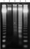
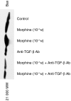
Laboratory and clinical reports indicate that opiate addicts are prone to infections. This effect of opiates is partly attributed to opiate-induced macrophage (Mphi) apoptosis. In the present study, we evaluated the role of transforming growth factor-beta (TGF-beta) in morphine-induced apoptosis of murine J774 cells and peritoneal Mphi. Mphi harvested from morphine-treated mice showed greater (P < 0. 0001) apoptosis when compared with control Mphi. Morphine also enhanced apoptosis of J774 cells and peritoneal Mphi. Anti-TGF-beta antibody inhibited (P < 0.001) the morphine-induced apoptosis in J774 cells (control 0.7 +/- 0.4%; 10-6 M morphine 23.5 +/- 0.7%; anti-TGF-beta antibody (Ab) + 10-6 M morphine 8.1 +/- 0.7%; apoptotic cells/field) and peritoneal Mphi (control 1.5 +/- 0.9%; 10-6 M morphine 29.1 +/- 1.4%; 10-6 M morphine + anti-TGF-beta Ab 19. 1 +/- 1.8%; apoptotic cells/field). TGF-beta enhanced (P < 0.001) apoptosis of J774 cells and peritoneal Mphi. TGF-beta also promoted Mphi DNA fragmentation into integer multiples of 180 bp (ladder pattern). Immunocytochemical studies revealed that morphine enhanced the Mphi cytoplasmic content of TGF-beta. In addition, Western blotting showed increased production of TGF-beta by morphine-treated J774 cells when compared with control cells. Morphine increased J774 cell expression of bax. Interestingly, morphine-induced bax expression was inhibited by anti-TGF-beta Ab. As both morphine-induced J774 cell apoptosis and bax expression were inhibited by anti-TGF-beta Ab, it appears that morphine-induced J774 cell apoptosis may be mediated through the generation of TGF-beta.
Figures


Similar articles
-
Morphine enhances macrophage apoptosis.J Immunol. 1998 Feb 15;160(4):1886-93. J Immunol. 1998. PMID: 9469450
-
Role of p38 mitogen-activated protein kinase phosphorylation and Fas-Fas ligand interaction in morphine-induced macrophage apoptosis.J Immunol. 2002 Apr 15;168(8):4025-33. doi: 10.4049/jimmunol.168.8.4025. J Immunol. 2002. PMID: 11937560
-
Ethanol-induced macrophage apoptosis: the role of TGF-beta.J Immunol. 1999 Mar 1;162(5):3031-6. J Immunol. 1999. PMID: 10072555
-
Morphine-induced macrophage apoptosis: oxidative stress and strategies for modulation.J Leukoc Biol. 2004 Jun;75(6):1131-8. doi: 10.1189/jlb.1203639. Epub 2004 Mar 23. J Leukoc Biol. 2004. PMID: 15039469
-
Morphine-induced macrophage apoptosis modulates migration of macrophages: use of in vitro model of urinary tract infection.J Endourol. 2002 Oct;16(8):605-10. doi: 10.1089/089277902320913314. J Endourol. 2002. PMID: 12470470
Cited by
-
Chronic opioid therapy in long-term cancer survivors.Clin Transl Oncol. 2017 Feb;19(2):236-250. doi: 10.1007/s12094-016-1529-6. Epub 2016 Jul 21. Clin Transl Oncol. 2017. PMID: 27443415 Review.
-
Modulation of immune function by morphine: implications for susceptibility to infection.J Neuroimmune Pharmacol. 2006 Mar;1(1):77-89. doi: 10.1007/s11481-005-9009-8. J Neuroimmune Pharmacol. 2006. PMID: 18040793 Review. No abstract available.
-
Restoration of Cyclo-Gly-Pro-induced salivary hyposecretion and submandibular composition by naloxone in mice.PLoS One. 2020 Mar 10;15(3):e0229761. doi: 10.1371/journal.pone.0229761. eCollection 2020. PLoS One. 2020. PMID: 32155179 Free PMC article.
-
The Effects of Opium Addiction on the Immune System Function in Patients with Fungal Infection.Addict Health. 2016 Fall;8(4):218-226. Addict Health. 2016. PMID: 28819552 Free PMC article.
-
Effects of Different Concentrations of Opium on the Secretion of Interleukin-6, Interferon-γ and Transforming Growth Factor Beta Cytokines from Jurkat Cells.Addict Health. 2015 Winter-Spring;7(1-2):47-53. Addict Health. 2015. PMID: 26322210 Free PMC article.
References
-
- Unanue ER. Macrophages, antigen-presenting cells and the phenomenon of antigen handling and presentation. In: Paul WE, editor. Fundamental Immunology. New York: Raven Press; 1989. p. 95.
-
- Szabo I, Rojavin M, Bussiere JL, et al. Suppression of peritoneal macrophage phagocytosis of Candida albicans by opioids. J Pharmacol Exp Ther. 1990;267:703. - PubMed
-
- Rojavin M, Szabo I, Bussiere JI, et al. Morphine treatment in vitro or in vivo decreases phagocytic function of murine macrophages. Life Sci. 1993;53:997. - PubMed
-
- Singhal PC, Pan C, Gibbons N. Effect of morphine on uptake of immunoglobulin G complexes by mesangial cells and macrophages. Am J Physiol. 1993;264:F859. - PubMed
-
- Tubaro E, Borelli G, Croce C, et al. Effect of morphine on resistance to infection. J Infect Dis. 1983;148:656. - PubMed
Publication types
MeSH terms
Substances
Grants and funding
LinkOut - more resources
Full Text Sources
Research Materials

