. 2000 May;100(1):70-6.
doi: 10.1046/j.1365-2567.2000.00008.x.

Expression of cytokine genes in Marek's disease virus-infected chickens and chicken embryo fibroblast cultures

Affiliations

Expression of cytokine genes in Marek's disease virus-infected chickens and chicken embryo fibroblast cultures

Z Xing et al. Immunology. 2000 May.

Abstract

The role of cytokines in the pathogenesis and immunity of Marek's disease (MD), a herpesvirus-induced T-cell lymphoma in chickens, is poorly understood. Two different experiments were used to examine the potential role of particular cytokines in the pathogenesis and immune responses of MD. First, chicken embryo fibroblasts (CEF) were stimulated with lipopolysaccharide (LPS) and/or recombinant chicken interferon-gamma (rChIFN-gamma) and used to develop techniques for examining transcription of IFN-alpha, IFN-gamma, inducible nitric oxide synthase (iNOS), interleukin (IL)-1beta, IL-2, IL-6 and IL-8 by reverse transcription-polymerase chain reaction (RT-PCR). Addition of LPS and/or rChIFN-gamma resulted in the up-regulation of mRNA for iNOS, IL-1beta and IL-6, while IFN-gamma was up-regulated by LPS alone. IL-2 was down-regulated by the treatments. Second, to determine the effects of Marek's disease herpesvirus (MDV) infection on cytokine transcription in vivo, chickens were infected with MDV at 21 days of age and examined at 7 days post-infection (p.i.) (exp. 1) or were infected with MDV at 1 day of age and examined from 3 to 15 days p.i. (exp. 2). In MDV-infected chickens, IFN-gamma transcription was up-regulated as early as 3 days p.i. until the termination of the experiment at 15 days p.i., while iNOS and IL-1beta were up-regulated between 6 and 15 days p.i. Infection of 1-day-old chicks increased levels of mRNA for IFN-gamma and iNOS between 16- and 64-fold at 9 days p.i. These results suggest that IFN-gamma and iNOS may play an important role in the pathogenesis of MD.


Figures
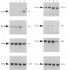
Figure 1
Figure 1
Expression of selected cytokine genes in chick embryo fibroblasts (CEF) stimulated with interferon-γ (IFN-γ) and lipopolysaccharide (LPS). Total RNAs were prepared from 5 × 10⁶ CEF cells 18 hr post-treatment with 50 units/ml of recombinant chicken IFN-γ (rChIFN-γ) (lane 1), 25 ng/ml of LPS (lane 2), 50 units/ml of rChIFN-γ+ 25 ng/ml of LPS (lane 3) or control (lane 4). Total RNAs were reverse transcribed using oligo-dT(16) as primers. cDNAs were amplified using primers specific for interleukin (IL)-2, IL-6, IL-8, IFN-α, IFN-γ, inducible nitric oxide synthase (iNOS) and β-actin, and a ‘touch-down’ polymerase chain reaction (PCR) procedure. PCR products were separated in 1·5% agarose gels, stained with ethidium bromide, visualized on the Eagle EyeII Still Video System (Stratagene) and photographed as negative images. The sizes of the respective PCR fragments are indicated by arrows.
Figure 2
Figure 2
Expression of interleukin-1β (IL-1β) in chick embryo fibroblasts (CEF) stimulated with interferon-γ (IFN-γ) and lipopolysaccharide (LPS). Total RNAs were prepared from 5 × 10⁶ CEF cells 18 hr post-treatment with 50 units/ml of recombinant chicken IFN-γ (rChIFN-γ) (lane 1), 25 ng/ml of LPS (lane 2), 50 units/ml of rChIFN-γ+ 25 ng/ml of LPS (lane 3) or control (lane 4). Total RNA was reverse transcribed with oligo dT(16) as primers. cDNA was amplified using primers specific for IL-1β and β-actin, using a ‘touch-down’ polymerase chain reaction (PCR) procedure. PCR products were separated in 1·5% agarose, stained with ethidium bromide, visualized on the Eagle EyeII Still Video System (Stratagene) and photographed as positive images. M, molecular weight ladder.
Figure 3
Figure 3
Cytokine expression in Marek's disease herpesvirus (MDV)-infected chicken splenocytes. Total RNAs were prepared from spleens prepared from 3-week-old control chickens (lane 1) and from chickens infected with JM16 (lane 2), SB-1 (lane 3) and HVT (lane 4), respectively. The RNAs were reverse transcribed using oligo dT(16) as primers and the cDNAs were amplified by the polymerase chain reaction (PCR) using specific primers for interferon-γ (IFN-γ), inducible nitric oxide synthase (iNOS), interleukin (IL)-2, IL-6 and IL-8. β-actin was used as a control. The amplified DNA fragments were analysed in a 1·5% gel, stained with ethidium bromide and visualized on the Eagle EyeII Still Video System (Stratagene). All images were photographed as negative images except for IL-8. The sizes of the PCR fragments or expected fragments (IL-6) are indicated.
Figure 4
Figure 4
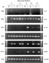
Temporal expression of cytokines in N2a chickens infected at 1 day of age with Marek's disease herpesvirus (MDV) strain JM16/19. Total RNA was prepared from pools of three spleens obtained from non-infected (N) and JM16/19-infected (I) chickens on 3, 6, 9, 12 and 15 days post-infection (p.i.) and used for reverse transcription–polymerase chain reaction (RT–PCR) analysis. Each lane represents a pool of three spleens obtained from different chickens. Primers specific for chicken interleukin (IL)-1β, IL-2, IL-6, IL-8, interferon-γ (IFN-γ), inducible nitric oxide synthase (iNOS) and β-actin were used. PCR products were electrophoresed on 1·5% agarose, stained with ethidium bromide and visualized on the Eagle EyeII Still Video System (Stratagene), and photographed as positive images. DNA markers (123-bp ladder) were used at both sides of the gel and the sizes of the respective cytokine gene fragments are indicated.
Figure 5
Figure 5
Comparison of expression of interferon-γ (IFN-γ) in a reverse transcription–competitive polymerase chain reaction (RT–cPCR) assay. cDNAs were generated from total RNAs prepared from JM-16-infected chickens at 9 days p.i. (a) or non-infected chickens (b) and incubated with fourfold dilutions of the multi synthetic standard (MSS) from 250 pg/μl (lane 7) to 0·06 pg/μl (lane 1) in the presence of 100 nm IFN-γ-specific primers. The cDNAs were amplified using a ‘touch-down’ PCR. The PCR products were separated by electrophoresis in 1·5% agarose gels, stained with ethidium bromide, visualized on the Eagle EyeII Still Video System (Stratagene) and photographed as negative images.
Figure 6
Figure 6
Comparison of expression of inducible nitric oxide synthase (iNOS) in a reverse transcription–competitive polymerase chain reaction (RT–cPCR) assay. cDNAs were generated from total RNAs prepared from JM-16-infected at 9 days p.i. (a) or non-infected chickens (b) and incubated with fourfold dilutions of the multi synthetic standard (MSS) from 250 pg/μl (lane 7) to 0·06 pg/μl (lane 1) in the presence of 100 nm iNOS-specific primers. The cDNAs were amplified using a ‘touch-down’ PCR. The PCR products were separated by electrophoresis in 1·5% agarose gels, stained with ethidium bromide, visualized on the Eagle EyeII Still Video System (Stratagene) and photographed as negative images.

References

    1. Schat KA, Chen C-LH, Calnek BW, Char D. Transformation of T-lymphocyte subsets by Marek's disease herpesvirus. J Virol. 1991;65:1408. - PMC - PubMed
    1. Calnek BW. Marek's disease – a model for herpesvirus oncology. CRC Crit Rev Microbiol. 1986;12:293. - PubMed
    1. Schat KA. Marek's disease: a model for protection against herpesvirus-induced tumors. Cancer Surveys. 1987;6:1. - PubMed
    1. Nazerian K, Lee LF, Yanagida N, Ogawa R. Protection against Marek's disease by a fowlpox virus recombinant expressing the glycoprotein B of Marek's disease virus. J Virol. 1992;66:1409. - PMC - PubMed
    1. Omar AR, Schat KA. Syngeneic Marek's disease virus (MDV)-specific cell-mediated immune responses against immediate early, late and unique MDV proteins. Virology. 1996;222:87. - PubMed

Publication types

LinkOut - more resources