Review
. 2000 May;10(5):624-33.
doi: 10.1101/gr.10.5.624.

Comparative genome mapping in the sequence-based era: early experience with human chromosome 7

Affiliations
Review

Comparative genome mapping in the sequence-based era: early experience with human chromosome 7

J W Thomas et al. Genome Res. 2000 May.

Abstract

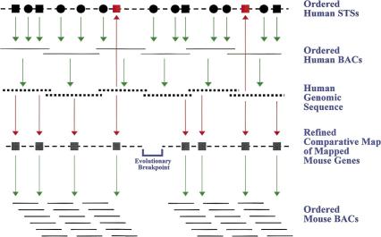
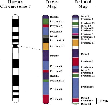
The success of the ongoing Human Genome Project has resulted in accelerated plans for completing the human genome sequence and the earlier-than-anticipated initiation of efforts to sequence the mouse genome. As a complement to these efforts, we are utilizing the available human sequence to refine human-mouse comparative maps and to assemble sequence-ready mouse physical maps. Here we describe how the first glimpses of genomic sequence from human chromosome 7 are directly facilitating these activities. Specifically, we are actively enhancing the available human-mouse comparative map by analyzing human chromosome 7 sequence for the presence of orthologs of mapped mouse genes. Such orthologs can then be precisely positioned relative to mapped human STSs and other genes. The chromosome 7 sequence generated to date has allowed us to more than double the number of genes that can be placed on the comparative map. The latter effort reveals that human chromosome 7 is represented by at least 20 orthologous segments of DNA in the mouse genome. A second component of our program involves systematically analyzing the evolving human chromosome 7 sequence for the presence of matching mouse genes and expressed-sequence tags (ESTs). Mouse-specific hybridization probes are designed from such sequences and used to screen a mouse bacterial artificial chromosome (BAC) library, with the resulting data used to assemble BAC contigs based on probe-content data. Nascent contigs are then expanded using probes derived from newly generated BAC-end sequences. This approach produces BAC-based sequence-ready maps that are known to contain a gene(s) and are homologous to segments of the human genome for which sequence is already available. Our ongoing efforts have thus far resulted in the isolation and mapping of >3,800 mouse BACs, which have been assembled into >100 contigs. These contigs include >250 genes and represent approximately 40% of the mouse genome that is homologous to human chromosome 7. Together, these approaches illustrate how the availability of genomic sequence directly facilitates studies in comparative genomics and genome evolution.


Figures

Figure 1
Figure 1
Overview of strategy for using human genomic sequence to facilitate human-mouse comparative mapping. Mapped and ordered human STSs (circles and squares correspond to random sequences and genes/ESTs, respectively) are used to isolate overlapping sets of human BACs, which in turn are sequenced. The resulting human genomic sequence can be readily aligned with the STS map by the electronic detection of mapped STSs. Also detected in the human sequence are previously unmapped sequences (e.g., genes/ESTs; depicted in red), thereby yielding an even more detailed STS map. Traditional comparative mapping can be enhanced with the human genomic sequence, specifically by the electronic detection of previously mapped mouse sequences (most often genes/ESTs). This allows refined comparative maps to be constructed that are more detailed than the starting human STS maps. The resulting linear order of markers on the comparative map allows more precise localization of evolutionary breakpoints at the ends of conserved segments. Finally, orthologous mouse sequences can be used to isolate corresponding mouse BACs and to assemble clone contigs. Red arrows reflect steps involving electronic analyses only, while green arrows reflect steps involving laboratory-based experimental analyses.
Figure 2
Figure 2
Small region of the refined human-mouse comparative map corresponding to a segment of human chromosome 7. On the left is a cytogenetic human-mouse comparative map of human chromosome 7q11-q22, based on information derived from MGI (ftp://ftp.informatics.jax.org/pub/informatics/reports/HMD_Human1.sq1.rpt).The predicted order of many of the same genes within this region of chromosome 7, as depicted on the Davis Human/Mouse Homology Map (DeBry & Seldin 1996) (see http://www.ncbi.nlm.nih.gov/Homology), is shown in the middle. On the right is our refined comparative map of the identical region, assembled with the aid of an available STS map (Bouffard et al. 1997) and genomic sequence from human chromosome 7 (see text for details). The complete refined comparative map is available at http://genome.nhgri.nih.gov/chr7/comparative. Note that the MGI-derived map includes a broader region than that shown for the Davis and refined maps. No definitive human gene order can be readily deduced from the crude cytogenetic positions available from the MGI-derived map. Using older information, the Davis map combined human cytogenetic mapping data with mouse genetic mapping data to deduce an order for human genes. All three maps indicate that this region of human chromosome 7 is orthologous to at least three different regions of the mouse genome. However, comparison of the Davis and refined maps reveals that the order of the proximal MMU6 and proximal MMU5 segments are inverted, the gene order within these segments is significantly different, and two genes included in the Davis map (ICA1 and RELN) are not present in this region. In general, human gene symbols are shown, except in cases where the human gene has not been named, in which case the mouse symbol or accession number is used. Lines connect identical genes present on both the Davis and refined maps. Genes depicted in bold are unique to one map, while those depicted in green on the MGI-derived map are located on MMU12.
Figure 3
Figure 3
Overview of human-mouse comparative maps for human chromosome 7. The refined comparative map of human chromosome 7 (see text for details) is shown on the right, along with the corresponding Davis Human/Mouse Homology Map (DeBry & Seldin 1996) (see http://www.ncbi.nlm.nih.gov/Homology) in the middle, and the chromosome 7 cytogenetic map on the left. The Davis map contains 14 orthologous segments from seven regions of the mouse genome. The refined map contains 20 orthologous segments from ten regions of the mouse genome. Evidence for all but one segment, MMU9 (which was defined by new genetic mapping data; J.W. Thomas and E.D. Green, unpubl.), is present in the MGI database. The refined map is drawn to scale, with the size of each established orthologous segment (in color) or unassigned region (white) estimated based on the number of mapped human STSs within the interval [assuming an average inter-STS spacing of 79 kb (Bouffard et al. 1997)]. The sizes of the conserved segments in the Davis map are not drawn to scale.
Figure 4
Figure 4
Accelerated construction of a mouse sequence-ready BAC contig using human genomic sequence. Roughly 180 kb of genomic sequence (top line) from human chromosome 7q22 (GenBank AC004022 and AC005021) was analyzed by RepeatMasker (A.F.A. Smit and P. Green, unpubl.; see http://www.genome.washington.edu/UWGC/analysistools/repeatmask.htm) to mask repetitive elements and then compared to GenBank using PowerBLAST (Zhang & Madden 1997). The small red dots below the human sequence represent a simplified view of matching mouse and rat mRNA/EST sequences. Three complete genes (PON1, PON2, PON3) and the 3′ end of a gene similar to rat neurabin (GenBank U72994) were detected in the region, with arrows indicating the direction of transcription. Based on this analysis, four overgo-type hybridization probes were designed from the matching mouse sequences (indicated by dashed arrows, with their names reflecting the corresponding GenBank accession numbers), optimizing for gene content and spacing (see text for details). These probes along with others designed from flanking human sequence were used to screen the mouse RPCI-23 BAC library, with the resulting probe-content data allowing assembly of three nascent contigs. The subsequent development and mapping of BAC insert end-specific overgo probes (indicated by squares) allowed the merger of the three contigs into the depicted >1-Mb contig from mouse chromosome 6.

References

    1. Battey J, Jordan E, Cox D, Dove W. An action plan for mouse genomics. Nat Genet. 1999;21:73–75. - PubMed
    1. Bouffard GG, Idol J R, Braden VV, Iyer LM, Cunningham AF, Weintraub LA, Touchman JW, Mohr-Tidwell RM, Peluso DC, Fulton RS, et al. A physical map of human chromosome 7: An integrated YAC contig map with average STS spacing of 79 kb. Genome Res. 1997;7:673–692. - PubMed
    1. Carver EA, Stubbs L. Zooming in on the human-mouse comparative map: genome conservation re-examined on a high-resolution scale. Genome Res. 1997;7:1123–1137. - PubMed
    1. Collins FS, Patrinos A, Jordan E, Chakravarti A, Gesteland R, Walters L Members of the DOE and NIH Planning Groups. New goals for the U.S. Human Genome Project: 1998–2003. Science. 1998;282:682–689. - PubMed
    1. Copeland NG, Jenkins NA, Gilbert DJ, Eppig JT, Maltais LJ, Miller JC, Dietrich WF, Weaver A, Lincoln SE, Steen RG, et al. A genetic linkage map of the mouse: current applications and future prospects. Science. 1993;262:57–66. - PubMed

LinkOut - more resources