. 2000 May;12(5):721-38.
doi: 10.1105/tpc.12.5.721.

Transgenic Arabidopsis plants expressing a fungal cutinase show alterations in the structure and properties of the cuticle and postgenital organ fusions

Affiliations

Transgenic Arabidopsis plants expressing a fungal cutinase show alterations in the structure and properties of the cuticle and postgenital organ fusions

P Sieber et al. Plant Cell. 2000 May.

Abstract

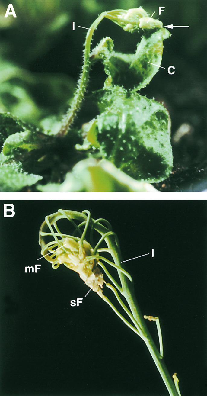
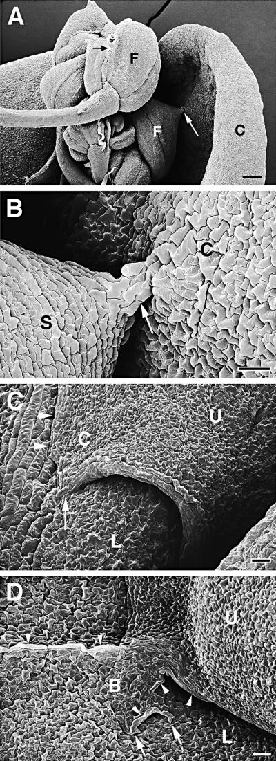
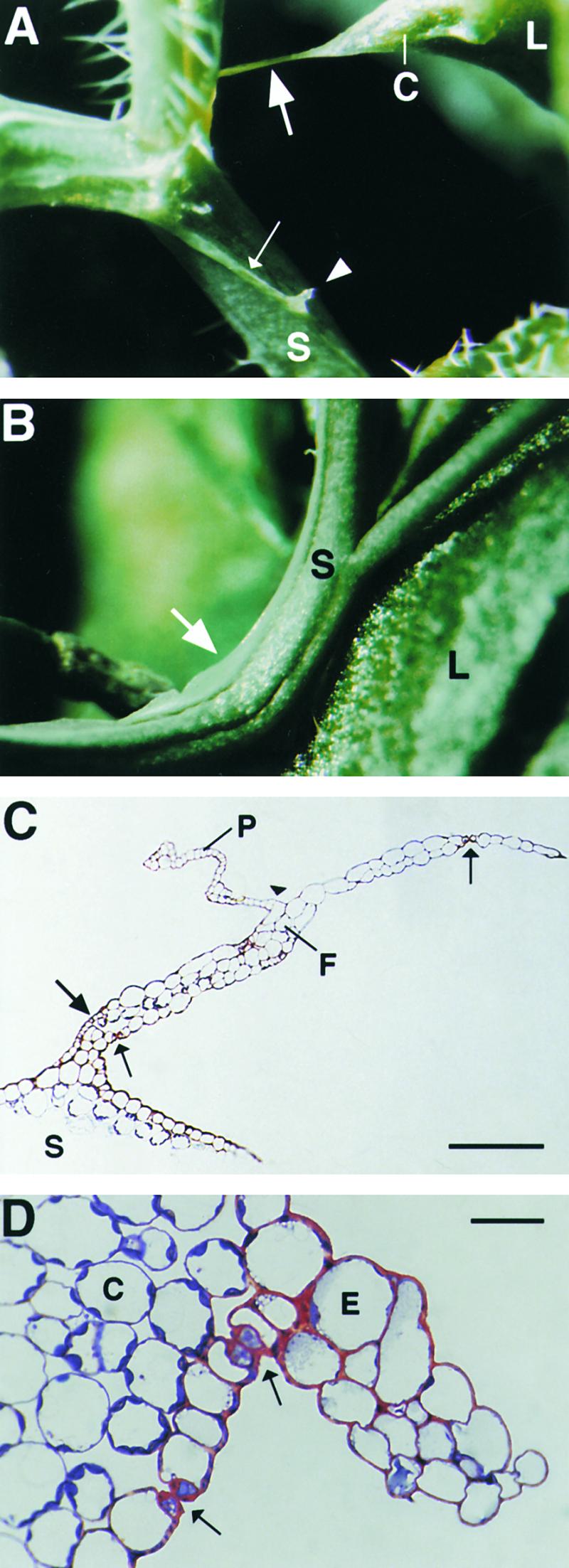
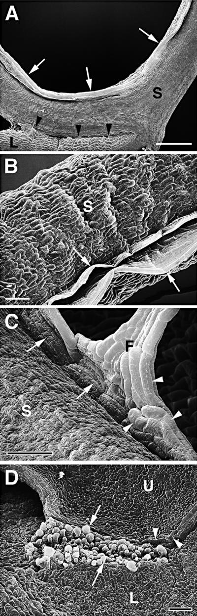
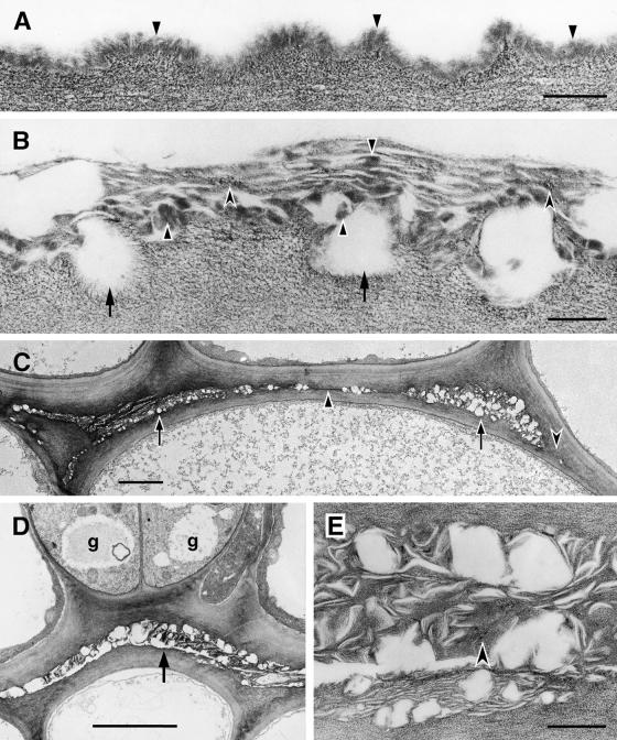
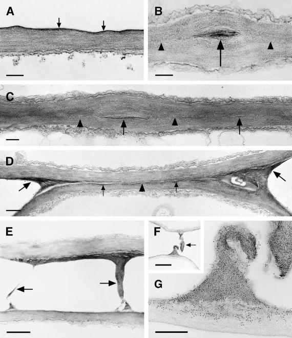
A major structural component of the cuticle of plants is cutin. Analysis of the function of cutin in vivo has been limited because no mutants with specific defects in cutin have been characterized. Therefore, transgenic Arabidopsis plants were generated that express and secrete a cutinase from Fusarium solani f sp pisi. Arabidopsis plants expressing the cutinase in the extracellular space showed an altered ultrastructure of the cuticle and an enhanced permeability of the cuticle to solutes. In addition, pollen could germinate on fully differentiated leaves of cutinase-expressing plants but not on control leaves. These differences coincided with strong postgenital organ fusions. The junctions of the fusions contained pectic polysaccharides. As fused organs grew apart from each other, organ deformations and protrusions of epidermal cells developed at positions with high mechanical stress. These results demonstrate that an intact cutin layer not only is important for plant-environment interactions but also prevents fusions between different plant organs and is therefore necessary for normal epidermal differentiation and organ formation.


Figures

Figure 1.
Figure 1.
Cutinase Activity of Transgenic Arabidopsis Expressing a Fungal Cutinase. Shown are nine representative transgenic Arabidopsis Col-0/gl1 lines transformed with the plasmid p35S::SS::Cut compared with Arabidopsis Col-0/gl1 control plants (C) transformed with the empty vector. T-1 to T-9, independent transformed plant lines 1 to 9.
Figure 2.
Figure 2.
Chlorophyll Release from Submerged Leaves of Col-0/gl1 Control Plants and Cutinase-Expressing Arabidopsis Plants. Open squares, wild-type Col-0/gl1 with a mean value from three analyses (±se); solid circles, diamonds, and triangles, representative measurement from cutinase-expressing Arabidopsis lines T-2, T-9, and T-5, respectively. The measurement was repeated three times with similar results. FW, fresh weight.
Figure 3.
Figure 3.
Pollen Germination on Rosette Leaves of Col-0/gl1 Control Plants and Cutinase-Expressing Arabidopsis Plants. (A) Mature leaf of a Col-0/gl1 plant fixed 20 hr after pollen application, stained with aniline blue, and visualized by UV excitation at 340 to 380 nm. The absence of green fluorescence demonstrates that no callose was deposited in the pollen and that no pollen tubes rich in callose developed. (B) Leaf of cutinase-expressing plant (line T-5) fixed 20 hr after pollen application, stained with aniline blue, and visualized by UV excitation at 340 to 380 nm. The green fluorescence indicates callose in pollen and particularly in outgrowing pollen tubes (Pt). In addition, callose was deposited in epidermal cells (E) underneath the germinating pollen as a defense response to pollen tubes penetrating the cuticle. formula image.
Figure 4.
Figure 4.
Visual Phenotype of Cutinase-Expressing Arabidopsis Plants. (A) Cutinase-expressing plant with a fusion between the inflorescence and a cauline leaf. The point of fusion is marked by an arrow. (B) Inflorescence of a cutinase-expressing plant with multiple organ fusion events between a series of flowers of different ages and among flower organs, leading to sterility. The points of cell contacts are hidden. C, cauline leaf; F, flower; I, inflorescence; mF, mature flower; sF, senescing flower.
Figure 5.
Figure 5.
Scanning Electron Microscopy of Organ Fusions of Cutinase-Expressing Arabidopsis Plants. (A) Five flowers with fused flower organs were fused to each other, and one flower was fused to a cauline leaf. Cell contacts between two sepals of one flower (small black arrow) and between a sepal and the cauline leaf (large white arrow) were visible. C, cauline leaf; F, flower. formula image. (B) Enlargement of (A). Small cell bridge (arrow) between sepal and cauline leaf. Epidermal cells appeared slightly larger in the cell bridge than in the unfused tissue. C, cauline leaf; S, sepal. formula image. (C) Two overlapping rosette leaves were fused to each other. The normally oval upper leaf formed a corner in the area of fusion. Suture with hidden cell contacts is indicated with arrowheads; visible cell contacts are indicated with an arrow. C, leaf corner; L, lower leaf; U, upper leaf. formula image. (D) A large cell bridge formed between two fused leaves. Large parts of the suture were not visible. Several cell contacts are visible (arrows). In part of the cell bridge, large epidermal cells developed (arrowheads), whereas the epidermal cell layer of other areas was normally differentiated. B, cell bridge; L, lower leaf; U, upper leaf. formula image.
Figure 6.
Figure 6.
Unusual Epidermal Protrusions Formed in Cutinase-Expressing Arabidopsis. (A) A pointed corner with a long epidermal attachment filament (large arrow) developed on a leaf after organ fusion with a stem under high mechanical stress. Epidermal ridges on a distorted stem are indicated by a small arrow; a protrusion originating from the ridge is marked by an arrowhead. C, leaf corner; L, leaf; S, stem. (B) Epidermal protrusion in a bent stem segment (arrow). L, leaf; S, stem. (C) Semithin section through an attachment filament. The large arrow indicates an area of high variability in size of epidermal cells; the small arrows indicate the stomata; and the arrowhead indicates the periclinal division plane. F, filament; P, protrusion; S, stem. Bar = 50 μm. (D) Semithin section through a ridge protruding from a distorted stem segment. Arrows indicate the stomata. C, cortical cell; E, epidermal cell. formula image.
Figure 7.
Figure 7.
Scanning Electron Micrographs of Epidermal Protrusions and Unusual Fusion Sutures of Cutinase-Expressing Arabidopsis Plants. (A) Epidermal protruding ridge (large arrows) in a bent stem segment and fusion between the stem and a leaf (arrowheads) at the distal side of the protrusion. formula image. (B) Specimen from (A) tilted and enlarged to visualize the two parallel protruding ridges (arrows) along the stem. formula image. (C) The base of a filament similar to the filament shown in Figure 6A that formed at an epidermal ridge (arrows) protruding along a stem. Of interest are the large cells in the protrusion (arrowheads). formula image. (D) Organ fusion between two leaves, leading to breakage of the epidermis. At the wound site, mesophyll cells bulged out from the inside of the leaf (arrows). Large tube-like cells lay within the plane of the epidermis close to the suture (arrowheads). formula image. F, filament; L, leaf; S, stem; U, upper leaf.
Figure 8.
Figure 8.
Ultrastructure of the Cuticle of the Stem Epidermis of Wild-Type and Cutinase-Expressing Arabidopsis Plants, and Organ Fusions between the Stems of a Transgenic Plant. (A) Col-0/gl1. Outer wall of an epidermal cell with cuticle of uniform structure (arrowheads) overlying the cell wall polysaccharides. formula image. (B) Cutinase-expressing Col-0/gl1. In contrast to the control, the contact zone between the cell wall polysaccharides and the cuticle was interrupted (arrows). Cuticular material accumulated in highly variable amounts, from apparently none to aggregates of mixed composition. Amorphous material of cuticular origin (solid arrowheads) was interspersed with polysaccharide microfibrils (concave arrowheads) in a loosely structured cuticle. formula image. (C) Cutinase-expressing Col-0/gl1. Fusion between two stems, overview. Apparently empty spaces surrounded by polysaccharidic cell wall materials characterize the fusion zone at most places (arrows). Areas containing small amounts of cuticular material can also be seen (solid arrowhead). At some places, the cuticle was interrupted and the cell walls of both epidermal cells came into direct contact (concave arrowhead). formula image. (D) Cutinase-expressing Col-0/gl1. Fusion between two stems. Two stomatal guard cells (g) were present in fused epidermal cell layers. Apparently empty spaces surrounded by polysaccharidic cell wall materials characterized this fusion zone (arrow). formula image. (E) Cutinase-expressing Col-0/gl1. Enlarged view of detail in (D). At this magnification, a large part of the materials in the fusion zone can be seen to have a fibrillar ultrastructure and to resemble cell wall polysaccharides (concave arrowhead). Waxy compounds were at least partially extracted during the dehydration and embedding procedures. formula image.
Figure 9.
Figure 9.
Ultrastructure of the Cuticle of the Leaf Epidermis, Structural Variations in the Fusion Zone between Leaves of Cutinase-Expressing Arabidopsis, and Identification of Pectic Polysaccharides as Essential Components of Organ Fusion. (A) Col-0/gl1. A thin electron-dense cuticle (arrows) overlays the cell wall polysaccharides in leaves. formula image. (B) and (C) Cutinase-expressing Col-0/gl1. The fusion zone between leaves is characterized by stretches of a direct contact of the polysaccharides (arrowheads) of the two epidermal cells and the local occurrence of interspersed cuticular material (arrows). Tilting the specimen (C) in the regions of a direct contact of polysaccharides confirmed the absence of detectable amounts of cuticular material or any structural change at the position where both cell walls came into contact (arrowheads). formula image. (D) Cutinase-expressing Col-0/gl1. The fusion zone is characterized by stretches of small amounts of cuticular material (small arrows) that is partially missing (arrowhead). In contrast to (C), the position at which the cell walls of both epidermal cells came into contact was still visible. At the positions of the cell corners where the fusion was tearing apart, electron-dense polysaccharide material accumulated (large arrows). formula image. (E) Stretched polysaccharide filaments (arrows) connecting two epidermal cells at points of high mechanical stress at the border of a fusion zone. formula image. (F) and (G) A large polysaccharide filament in a fusion zone probably originated under mechanical stress. (F) Overview of the region; the arrow indicates the polysaccharide filament. Bar = 2 μm. (G) Immunolabeling of the polysaccharides involved in organ fusion by JIM5 monoclonal antibodies, specific for pectin with low degrees of esterification. Bar = 500 nm.

References

    1. Baker, C.J., McCormick, S.L., and Batemann, D.F. (1982). Effects of purified cutin esterase upon the permeability and mechanical strength of cutin membranes. Phytopathology 72, 420–423.
    1. Baker, E.A. (1982). Chemistry and morphology of plant epicuticular waxes. In The Plant Cuticle, D.F. Cutler, K.L. Alvin, and C.E. Price, eds (London: Academic Press), pp. 139–162.
    1. Bechtold, N., Ellis, J., and Pelletier, G. (1993). In planta Agrobacterium-mediated gene transfer by infiltration of adult Arabidopsis thaliana plants. C. R. Acad. Sci. 316, 1194–1199. - PubMed
    1. Becraft, P.W., Stinard, P.S., and McCarty, D.R. (1996). CRINKLY4: A TNKR-like receptor kinase involved in epidermal maize differentiation. Science 273, 1406–1409. - PubMed
    1. Deising, H., Nicholson, R.L., Haug, M., Howard, R.J., and Mendgen, K. (1992). Adhesion pad formation and the involvement of cutinase and esterases in the attachment of uredospores to the host cuticle. Plant Cell 4, 1101–1111. - PMC - PubMed

Publication types

MeSH terms