. 2000 May;12(5):771-86.
doi: 10.1105/tpc.12.5.771.

Pti4 is induced by ethylene and salicylic acid, and its product is phosphorylated by the Pto kinase

Affiliations

Pti4 is induced by ethylene and salicylic acid, and its product is phosphorylated by the Pto kinase

Y Q Gu et al. Plant Cell. 2000 May.

Abstract

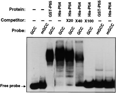
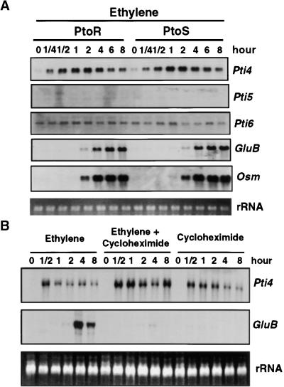
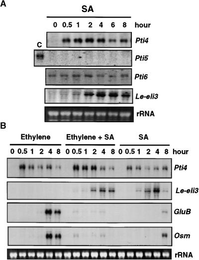
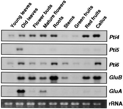
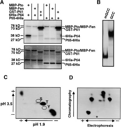
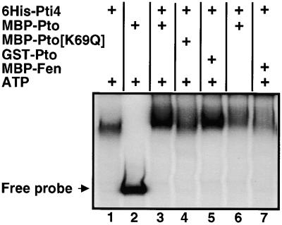
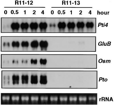
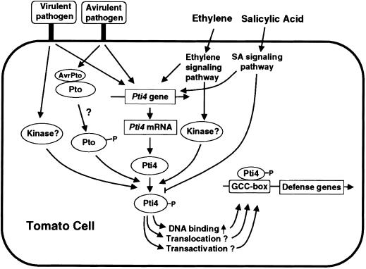
The tomato Pti4 gene encodes a transcription factor that was identified on the basis of its specific interaction with the product of the Pto disease resistance gene in a yeast two-hybrid system. We show here that the Pti4 protein specifically binds the GCC-box cis element, which is present in the promoter region of many pathogenesis-related (PR) genes. Expression of the Pti4 gene in tomato leaves was rapidly induced by ethylene and by infection with Pseudomonas syringae pv tomato, and this induction preceded expression of GCC-box-containing PR genes. Although salicylic acid also induced Pti4 gene expression, it did not induce GCC-box PR genes. Rather, salicylic acid antagonized ethylene-mediated expression of GCC-box PR genes. We demonstrate that the Pti4 protein is specifically phosphorylated by the Pto kinase and that this phosphorylation enhances binding of Pti4 to the GCC box. In addition, induced overexpression of Pto and Pti4 in tomato leaves resulted in a concomitant increase in GCC-box PR genes. Our results support a model in which phosphorylation of the Pti4 protein by the Pto kinase enhances the ability of Pti4 to activate expression of GCC-box PR genes in tomato.


Figures

Figure 1.
Figure 1.
Pti4 Specifically Binds the GCC-Box Sequence. A mobility shift assay was performed as described previously (Zhou et al., 1995), using 50 ng of purified His-Pti4 or GST-Pti5 fusion protein mixed with 5 fmol of 32P-labeled wild-type GCC-box or mGCC-box oligonucleotides (see Methods). The competitor consisted of the unlabeled wild-type GCC oligonucleotide. The notations ×20, ×40, and ×100 indicate that the amount of unlabeled wild-type GCC-box oligonucleotide added to these reactions was 20-, 40-, or 100-fold that of the labeled GCC-box oligonucleotide. The (−) signs indicate that no competitor, or no protein, was added to these reactions. The position of the unbound (Free) probe is shown.
Figure 2.
Figure 2.
Effect of Ethylene and Cycloheximide on Expression of Pti4, Pti5, Pti6, and GCC-Box-Containing PR Genes. Fully expanded leaves from 4-week-old tomato plants were used for analysis of gene expression. Equal loading of lanes in all gels was verified by visualizing the rRNA in the RNA gels stained with ethidium bromide. (A) RG-PtoR or RG-PtoS tomato plants were treated with 10 μL/L ethylene gas for the times indicated. Total RNAs were isolated from leaves and analyzed as duplicate RNA gel blots by hybridization with radiolabeled Pti4, Pti5, Pti6, GluB, or Osm probes. (B) RG-PtoR plants were treated with ethylene (10 μL/L), cycloheximide (5 μg/mL), or a combination of both for the times indicated. Total RNAs were isolated from leaves and analyzed as duplicate RNA gel blots by hybridization with radiolabeled Pti4 or GluB probes.
Figure 3.
Figure 3.
Effect of SA and Ethylene on Expression of Pti4, Pti5, Pti6, Le-eli3, and the GCC-Box-Containing PR Genes GluB and Osm. Fully expanded leaves from 4-week-old tomato plants were used for analysis of gene expression. Equal loading was verified by visualizing the rRNA in the RNA gels stained with ethidium bromide. (A) RG-PtoR tomato plants were treated with 1 mM SA for the times indicated. Total RNAs were isolated from leaves and analyzed as duplicate RNA gel blots by hybridization with radiolabeled Pti4, Pti5, Pti6, or Le-eli3 probes. Note that lane C for Pti5 contains total RNA isolated from leaves of 8-week-old tomato plants in which the Pti5 transcript is detectable. (B) RG-PtoR tomato plants were treated with ethylene (10 μL/L), SA (1 mM), or a combination of both for the times indicated. Total RNAs were isolated from leaves and analyzed as duplicate RNA gel blots by hybridization with radiolabeled Pti4, Le-eli3, GluB, or Osm probes.
Figure 4.
Figure 4.
Effect of Wounding or Systemin on Expression of Pti4, Pti5, Pti6, Pin2, and GluB. Fully expanded leaves from 4-week-old tomato plants were used for analysis of gene expression. Equal loading was verified by visualizing the rRNA in the RNA gels stained with ethidium bromide. Pin II, Pin2. (A) Tomato RG-PtoR plants were wounded by rubbing leaves with carborundum, after which total RNAs were isolated from leaves at the times indicated. Duplicate RNA gel blots were hybridized with radiolabeled Pti4, Pti5, Pti6, Pin2, or GluB probes. Note that lane C for Pti5 contains total RNA isolated from leaves of 8-week-old tomato plants in which Pti5 transcript is detectable. (B) Tomato RG-PtoR plants were treated with 1 nM systemin, after which total RNAs were isolated from leaves at the times indicated. Duplicated RNA gel blots were hybridized with radiolabeled Pti4 or Pin2 probes.
Figure 5.
Figure 5.
Expression of Pti4, Pti5, Pti6, GluB, and GluA in Various Tissues at Different Developmental Stages. The third and fourth leaves of 3- to 4-week-old or 11- to 12-week-old tomato plants were harvested as young and old leaves, respectively. Unopened and fully opened flowers are referred to as flower buds and mature flowers, respectively. Root and stem tissues were harvested from 3- to 4-week-old plants. Green fruits were immature fruits, and red fruits were red ripe fruits. Total RNAs were isolated from these tissues, and duplicate RNA gel blots were hybridized with the Pti4, Pti5, Pti6, GluB, or GluA probes. Equal loading was verified by visualizing the rRNA in the RNA gel stained with ethidium bromide.
Figure 6.
Figure 6.
Expression of Pti4, Pti6, GluB, and Osm in an Incompatible or Compatible Interaction Involving P. s. tomato. (A) RG-PtoR or RG-PtoS tomato plants were vacuum-infiltrated with 2.2 × 10⁷ colony-forming units per milliliter of P. s. tomato DC3000 expressing the avrPto gene in 10 mM MgCl2 and 0.005% Silwet. (B) RG-PtoR tomato plants were vacuum-infiltrated with 10 mM MgCl2 and 0.005% Silwet as a control. Leaf tissues were harvested at the indicated times after vacuum infiltration. Total RNAs were isolated, and duplicate RNA gel blots were hybridized with radiolabeled Pti4, Pti6, GluB, or Osm probes. Equal loading was verified by visualizing the rRNA in the RNA gels stained with ethidium bromide.
Figure 7.
Figure 7.
Phosphorylation of the Pti4 Protein by Pto Kinase. (A) His-Pti4, Pti5-His, MBP-Pto, MBP-Fen, and GST-Pti1 proteins were purified as described in Methods. Two micrograms of His-Pti4 or Pti5-His protein was incubated with 2 μg of MBP-Pto, MBP-Fen, or GST-Pti1 protein in an in vitro kinase assay. Total proteins from each reaction were separated by SDS-PAGE and analyzed by autoradiography. The autoradiograph (top) shows the phosphorylated proteins, and the Coomassie blue–stained gel (bottom) shows the protein profile. The (+) signs denote that the indicated protein was present in the reaction. Protein masses are shown in kilodaltons. (B) A mobility shift assay was performed as described previously (Zhou et al., 1995), using 50 ng of purified Pti5-His fusion protein mixed with 5 fmol of 32P-labeled wild-type GCC-box or mGCC-box oligonucleotides (see Methods). (C) Radiolabeled His-Pti4 protein that had been phosphorylated by Pto was transferred to a polyvinyl difluorine membrane, eluted, and hydrolyzed to its compositional amino acids as described in the Methods. The amino acids were separated by two-dimensional thin-layer chromatography. The (+) and (−) signs indicate the positions of the positive and negative electrodes. The locations of standards of serine (S), threonine (T), and tyrosine (Y) residues as determined by addition of ninhydrin are indicated. Radiolabeled, phosphorylated residues were detected by autoradiography. (D) Radiolabeled His-Pti4 protein that had been phosphorylated by Pto was digested with trypsin, as described by Sessa et al. (1998). Peptides were separated horizontally by thin-layer electrophoresis at pH 4.7 and then vertically by ascending chromatography. The (+) and (−) signs indicate the positions of the positive and negative electrodes. Peptides were detected by autoradiography. The origin is indicated by an asterisk.
Figure 8.
Figure 8.
Phosphorylation of Pti4 by Pto Affects Its DNA Binding Activity. Components as indicated in each lane were preincubated at 25°C for 30 min in 10 μL of kinase buffer. For each kinase reaction, 0.25 μg of His-Pti4 and kinase was used. The final concentration of unlabeled ATP was 1 mM. After incubation, the volume in each tube was adjusted to 30 μL, with the mobility shift assay buffer containing the radiolabeled probe, and assayed as described in Methods. The (+) signs indicate that the corresponding protein, or ATP, was present in the reaction. The position of the unbound (Free) probe is shown.
Figure 9.
Figure 9.
Expression of Pti4, the GCC-Box-Containing PR Genes, and Pto Induced by SA in Tomato Plants with or without a 35::Pto Transgene. A tomato line that is homozygous for a 35S::Pto transgene (R11-12) and a sibling line lacking the transgene (R11-13) were treated with 1 mM SA, and leaves were harvested at the times indicated. Total RNAs were isolated, and duplicate RNA gel blots were hybridized with radiolabeled Pti4, GluB, Osm, or Pto probes. Equal loading was verified by visualizing the rRNA in the RNA gel stained with ethidium bromide.
Figure 10.
Figure 10.
Proposed Model for Activation of GCC-Box-Containing PR Genes by Transcription Factor Pti4 in Pto-Mediated and Other Defense Response Signaling Pathways. ?, mechanistic steps that are still conjectural; P, phosphorylation.

Similar articles

Cited by

References

    1. Abeles, F.B., Morgan, P.W., and Saltveit, M.E., Jr. (1992). Ethylene in Plant Biology. (San Diego, CA: Academic Press).
    1. Alfano, J.R., and Collmer, A. (1996). Bacterial pathogens in plants: Life up against the wall. Plant Cell 8, 1683–1698. - PMC - PubMed
    1. Ausubel, F.M., Brent, R., Kingston, R.E., Moore, D.D., Smith, J.A., Seidman, J.G., and Struhl, K., eds (1987). Protocols in Molecular Biology. (New York: John Wiley).
    1. Broglie, K.E., Chet, I., Holliday, M., Cressman, R., Biddle, P., Knowlton, S., Mauvais, C.J., and Broglie, R. (1991). Transgenic plants with enhanced resistance to the fungal pathogen Rhizoctonia solani. Science 254, 1194–1197. - PubMed
    1. Buchel, A.S., Molemkamp, R., Bol, J.F., and Linthorst, H.J.M. (1996). The PR-1a promoter contains a number of elements that bind GT-1–like nuclear factors with different affinity. Plant Mol. Biol. 30, 493–504. - PubMed

Publication types

MeSH terms