. 2000 May;12(5):787-802.
doi: 10.1105/tpc.12.5.787.

Identification, purification, and molecular cloning of a putative plastidic glucose translocator

Affiliations

Identification, purification, and molecular cloning of a putative plastidic glucose translocator

A Weber et al. Plant Cell. 2000 May.

Abstract

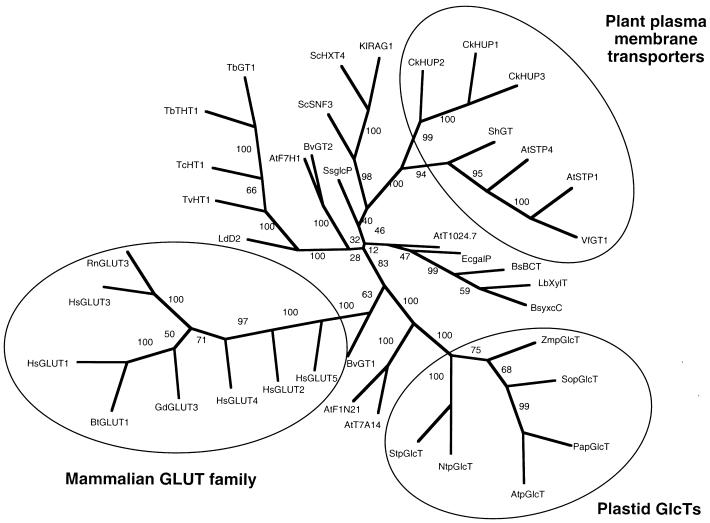
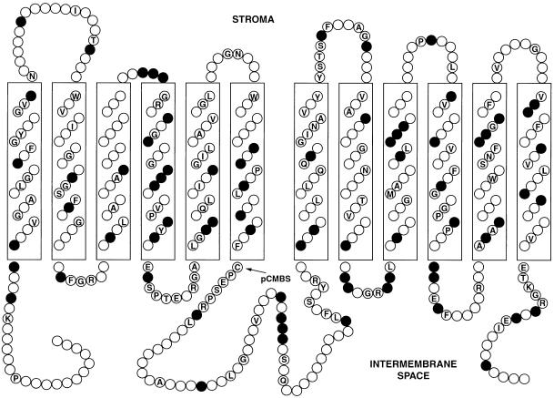
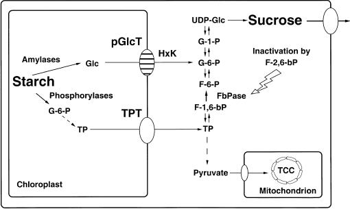
During photosynthesis, part of the fixed carbon is directed into the synthesis of transitory starch, which serves as an intermediate carbon storage facility in chloroplasts. This transitory starch is mobilized during the night. Increasing evidence indicates that the main route of starch breakdown proceeds by way of hydrolytic enzymes and results in glucose formation. This pathway requires a glucose translocator to mediate the export of glucose from the chloroplasts. We have reexamined the kinetic properties of the plastidic glucose translocator and, using a differential labeling procedure, have identified the glucose translocator as a component of the inner envelope membrane. Peptide sequence information derived from this protein was used to isolate cDNA clones encoding a putative plastidic glucose translocator from spinach, potato, tobacco, Arabidopsis, and maize. We also present the molecular characterization of a candidate for a hexose transporter of the plastid envelope membrane. This transporter, initially characterized more than 20 years ago, is closely related to the mammalian glucose transporter GLUT family and differs from all other plant hexose transporters that have been characterized to date.


Figures

Figure 1
Figure 1
Time Course of Uptake of 4 mM d-14C-Glc into Spinach Chloroplasts. Shown is the radioactivity present in neutral (closed circles) and acidic (open circles) products. Error bars indicate standard deviation. Chl, chlorophyll.
Figure 2
Figure 2
Determination of the Kinetic Constants of pGlcT. (A) Substrate dependency of uptake of d-14C-Glc into spinach chloroplasts. Chl, chlorophyll; h, hour. (B) Double-reciprocal representation (Lineweaver–Burk plot) of data shown in (A). Error bars indicate standard deviation. 1/v, reciprocal value of the Glc uptake rate; 1/mM, reciprocal value of the substrate concentration.
Figure 3
Figure 3
Effect of NEM Treatment on the Rate of Glc Transport into Spinach Chloroplasts. The effect of pretreatment of spinach chloroplasts with NEM in the presence of sorbitol or Glc on the resulting rate of 20 mM D-14C-Glc uptake is shown. Chloroplasts were incubated in buffer B and 1 mM DTT for 15 min at 20°C, collected, and resuspended three times to remove DTT. Aliquots were resuspended in an osmoticum containing either 330 mM sorbitol (open circles) or 330 mM Glc (closed circles) for 15 min at 20°C. NEM was added at the concentration shown, and incubation continued for another 30 min. Chloroplasts were collected and resuspended three times to remove NEM and Glc. Uptake of 4 mM D-14C-Glc was measured in 1-sec assays. Error bars indicate standard deviation. Chl, chlorophyll; h, hour.
Figure 4.
Figure 4.
Differential Labeling of pGlcT Using 3H- and 14C-NEM for Glc-Protected and Glc-Unprotected Envelope Proteins and Their Separation on SDS–Polyacrylamide Gels. Labeling of Glc-protected or Glc-unprotected spinach chloroplasts was performed as described in the text. Chloroplast envelopes were prepared, and membrane proteins were partitioned between n-butanol and water. Fractions were dried, taken up in sample buffer, and subjected to SDS-PAGE. After staining and drying, the gel was cut into 1-mm sections, and the tritium and 14C present in each section were determined. Data are plotted as the difference in percentage of total radioactivity present in each section (14Csection/14Ctotal3Hsection/3Htotal). At the bottom of each plot are the gels, showing the positions of proteins with respect to the sections cut from the gel. (A) n-Butanol fraction. (B) Aqueous fraction. (C) Envelope membranes were extracted by 20-fold excess n-butanol. The butanol fraction was dried, washed with n-hexane, and analyzed by SDS-PAGE. The 43-kD GlcT protein is indicated by an arrow.
Figure 5.
Figure 5.
Evolutionary Relationships of Hexose Transporters Represented as an Unrooted Phylogenetic Tree. Transporters belonging to the families of mammalian hexose transporters (GLUT family), plant plasma membrane transporters, and the plastidic GlcTs are encircled. The first two letters of the acronyms indicate the species (At, Arabidopsis thaliana; Bs, Bacillus subtilis; Bt, Bos taurus; Bv, Beta vulgaris; Ck, Chlorella kessleri; Ec, Escherichia coli; Gd, Gallus domesticus; Hs, Homo sapiens; Kl, Kluyveromyces lactis; Lb, Lactobacillus brevis; Ld, Leishmania donovani; Nt, Nicotiana tabacum; Pa, Prunus armeniaca; Rn, Rattus norwegicus; Sc, Saccharomyces cerevisiae; Sh, Saccharum hybrid cultivar; So, Spinacia oleracea; Ss, Synechocystis sp (PCC6803); St, Solanum tuberosum; Tb, Trypanosoma brucei; Tc, T. cruzi; Tv, T. vivax; Vf, Vicia faba; Zm, Zea mays. Bootstrap values are depicted on the respective branches.
Figure 6.
Figure 6.
Schematic Representation of the Proposed Transmembrane Topology of the Putative Mature pGlcT Protein from Spinach Chloroplast Envelope Membranes. The 12 transmembrane helices are shown as boxes. Identical amino acids in the human GLUT1 and the spinach chloroplast envelope membrane pGlcT are indicated by their single-letter abbreviations, and chemically similar residues (D,E; F,Y,W; I,L,V,M; K,R; N,Q; and S,T) are noted by shaded circles. The cysteine residue that is protected by Glc from labeling with pCMBS is indicated by an arrow. Adapted from Gould and Bell (1990).
Figure 7.
Figure 7.
Importation of the 35S-Labeled pGlcT Precursor Protein into Isolated Spinach Chloroplasts. Importation was performed at 25°C either in the dark (lane 2) or in the light (lane 1 and lanes 3 to 5) and in the presence of 2 mM ATP (lanes 4 and 5) or 5 μM carbonylcyanide m-chlorophenylhydrazone (lane 2). Sample 1 (lane 1) contained plastids that were pretreated with thermolysin (30 μg mL−1). After importation, sample 5 (lane 5) was further treated with thermolysin (50 μg mL−1) in the presence of 1 mM CaCl2 for 30 min. Envelope membranes were isolated as described earlier (Flügge et al., 1989) and analyzed by SDS-PAGE and phosphoimaging. p, i, and m represent the precursor protein, the intermediate form, and the mature form, respectively.
Figure 8.
Figure 8.
Expression Analysis of the Gene Encoding pGlcT. (A) RNA gel blot analysis of pGlcT mRNA in different tissues of tobacco. Thirty micrograms of total RNA isolated from source leaves, stems, sink leaves, roots, flower buds, and petioles was hybridized with the pGlcT cDNA probe from spinach after gel electrophoresis and subsequent transfer of the RNA to a nylon membrane. (B) Diurnal variation of the steady state concentration of pGlcT mRNA in source leaves of tobacco. Total RNA was isolated from source leaves of tobacco plants after 10 hr of light (lane 1), 1 hr of dark (lane 2), 4 hr of dark (lane 3), 12 hr of dark (lane 4), 1 hr of light (lane 5), and 3 hr of light (lane 6) during a 12-hr-light/12-hr-dark cycle. Analysis (30 μg per lane) was performed as indicated in (A). (C) RNA gel blot analysis of pGlcT and TPT mRNA of wild-type and transgenic tobacco plants. Total RNA was isolated from wild-type plants (lanes 1 and 2) and from plants showing an antisense repression of the chloroplastic TPT (lanes 3 to 6; Häusler et al., 1998). Analysis (50 μg per lane) was performed as in (A).
Figure 9.
Figure 9.
Proposed Function of the pGlcT Protein in Starch Mobilization. Glc from hydrolytic breakdown of transitory starch is exported from the chloroplasts by pGlcT and subsequently converted to Glc 6-phosphate (G-6-P) by a hexokinase (HxK) that is located in the chloroplast outer envelope membrane. Glucose 6-phosphate is shuttled to the site of sucrose biosynthesis, whereas trioseP that results from phosphorolytic starch breakdown is subjected to glycolysis. F-6-P, fructose 6-phosphate; F-1,6-bP, fructose 1,6-bisphosphate; F-2,6-bP, fructose 2,6-bisphosphate; FbPase, fructose 1,6-bisphosphate phosphatase; G-1-P, Glc 1-phosphate; TCC, tricarboxylic acid cycle; TP, trioseP.

References

    1. Baldwin, S.A., and Lienhard, G.E. (1989). Purification and reconstitution of glucose transporter from human erythrocytes. Methods Enzymol. 174, 39–50. - PubMed
    1. Bechtold, N., Ellis, G., and Pelletier, G. (1993). In planta Agrobacterium-mediated gene transfer by infiltration of adult Arabidopsis thaliana plants. C. R. Acad. Sci. 316, 1194–1199. - PubMed
    1. Beck, E. (1985). The degradation of transitory starch in chloroplasts. In Regulation of Carbon Partitioning in Photosynthetic Tissue, R.L. Heath and J. Preiss, eds (Baltimore, MD: Waverly), pp. 27–44.
    1. Capaldi, R.A., and Vanderkooi, G. (1972). The low polarity of many membrane proteins. Proc. Natl. Acad. Sci. USA 69, 930–932. - PMC - PubMed
    1. Caspar, T., Huber, S.C., and Somerville, C. (1985). Alterations in growth, photosynthesis, and respiration in a starchless mutant of Arabidopsis thaliana (L) deficient in chloroplast phosphoglucomutase activity. Plant Physiol. 79, 11–17. - PMC - PubMed

Publication types

MeSH terms