. 2000 May 15;19(10):2257-69.
doi: 10.1093/emboj/19.10.2257.

Thr38 and Ser198 are Pto autophosphorylation sites required for the AvrPto-Pto-mediated hypersensitive response

Affiliations

Thr38 and Ser198 are Pto autophosphorylation sites required for the AvrPto-Pto-mediated hypersensitive response

G Sessa et al. EMBO J. .

Erratum in

  • EMBO J 2000 Jun 15;19(12):3157

Abstract

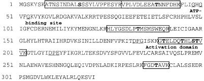
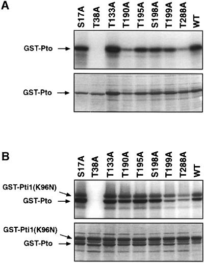
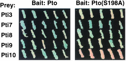
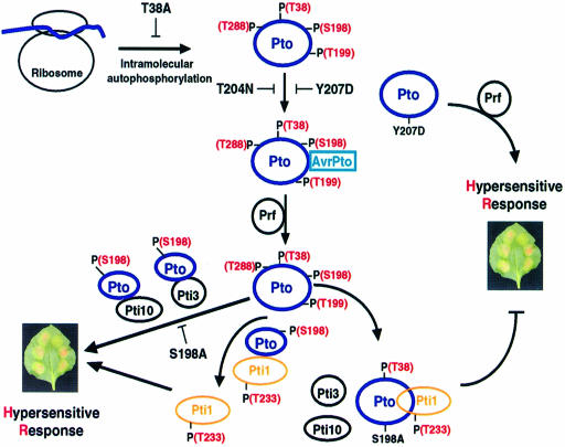
The tomato Pto kinase confers resistance to Pseudomonas syringae pv. tomato expressing the AvrPto protein. To elucidate the role of Pto autophosphorylation in disease resistance, eight sites autophosphorylated by Pto in vitro were identified by a combination of HPLC purification of tryptic phosphopeptides, MALDI-TOF/MS analysis and Edman degradation. Mutational analysis of the autophosphorylation sites revealed that Pto residues Thr38 and Ser198 are required for AvrPto-Pto- mediated elicitation of a hypersensitive response in the plant. Thr38, which is the main Pto autophosporylation site and is located outside the kinase catalytic domain, was also required for Pto kinase activity and its physical interaction with AvrPto, the Pti1 kinase and the transcription factor Pti4. Ser198, located in the Pto activation domain, was dispensable for kinase activity and for interaction with AvrPto. However, a mutation at this site resulted in altered Pto interactions with the Pti1 kinase and the Pto interactors of unknown function Pti3 and Pti10. These results suggest that autophosphorylation events at Pto Thr38 and Ser198 are required for signal transduction by Pto and participate in distinct molecular mechanisms.


Figures

None
Fig. 1. Reverse-phase HPLC fractionation of a tryptic digest of autophosphorylated GST–Pto. A tryptic digest of autophosphorylated GST–Pto was fractionated by reverse-phase HPLC using a C18 column. Peptides were eluted by a gradient of buffer B, containing 0.01% TFA, 75% acetonitrile and 25% water (dotted line). The radioactivity of each fraction was determined by Cerenkov counting (circles).
None
Fig. 2. Identification of Pto autophosphorylation sites. (A) MALDI-TOF/MS spectrum of HPLC-purified peak G, derived from a tryptic digest of autophosphorylated GST–Pto. m/z values for the major components of the spectrum, and the sequence of the phosphopeptide identified are indicated. (B) Reverse-phase HPLC fractionation of a tryptic digest of autophosphorylated GST–Pto(T288A). A tryptic digest of autophosphorylated GST–Pto(T288A) was fractionated by reverse-phase HPLC using a C18 column. Peptides were eluted by a gradient of buffer B, containing 0.01% TFA, 75% acetonitrile and 25% water (dotted line). The radioactivity of each fraction was determined by Cerenkov counting (circles). (C) Manual Edman degradation of phosphopeptide 8–28. The partially purified phosphopeptide 8–28, detected in peak I, was immobilized on a membrane disk and subjected to sequential cycles of Edman degradation. Bars represent the amount of radioactivity released after each cycle. The amino acid sequence of peptide 8–28 is shown on top of the diagram.
None
Fig. 3. Phosphopeptides and autophosphorylation sites identified in the Pto amino acid sequence by analysis of a GST–Pto tryptic digest. Sequences of tryptic phosphopeptides are boxed, and residues identified as autophosphorylation sites are in bold. Glycine residues of the conserved motif GXGXXG for the ATP-binding site, and the DFG and PE motifs representing the borders of the kinase activation domain are underlined (Hanks and Quinn, 1991).
None
Fig. 4. Elicitation of the hypersensitive response (HR) by expression of Pto autophosphorylation mutants and AvrPto in N.benthamiana leaves. (A) Transient expression assay of Pto(T38A) for the HR in N.benthamiana leaves. Mature leaves were infiltrated with A.tumefaciens EHA105 strains (OD600 = 0.4) that contained the pBTEX plasmid expressing the following combinations of proteins: in 1, 2 and 3, Pto(T38A) and AvrPto; in 4, AvrPto; and in 5 and 6, Pto and AvrPto. The HR was visualized as localized tissue collapse typically within 3–5 days. (B) Transient expression assay of Pto(S198A) for the HR in N.benthamiana leaves. Mature leaves were infiltrated as described in (A), with A.tumefaciens strains expressing the following combinations of proteins: in 1, 2 and 3, Pto(S198A) and AvrPto; in 4, AvrPto; and in 5 and 6, Pto and AvrPto. (C) An HR index, reflecting the efficiency of the indicated mutants in the elicitation of the HR in vivo, was calculated as described in Materials and methods. Calculation of the HR index included at least 82 independent observations for each mutant.
None
Fig. 5. In vitro kinase assay of Pto forms individually mutated at autophosphorylation sites. The effect of mutations at autophos phorylation sites on Pto autophosphorylation activity (A), and Pto phosphorylation of a kinase-deficient Pti1(K96N) (B), were tested in kinase assays in vitro. Pto mutants and Pti1(K96N) were expressed in bacteria as GST fusions. Kinase activity was tested in reactions containing the combinations of fusion proteins indicated in the figure. Proteins were fractionated by 10% SDS–PAGE and analyzed with a phosphoimager. The phosphoimager exposure (top panel) shows the proteins phosphorylated in each assay, whereas the Coomassie-stained gel (bottom panel) shows the protein species present in the reaction mixtures.
None
Fig. 6. Yeast two-hybrid interactions of Pto autophosphorylation sites mutants with AvrPto, Pti1 and Pti4. (A) Expression of bait fusion proteins in the yeast strain EGY48. Wild-type and mutant forms of Pto were expressed in yeast as LexA fusions in the bait plasmid pEG202. LexA fusions were detected by chemiluminescent visualization in Western blots using polyclonal antibodies raised against LexA. In each lane, the Pto form expressed in the yeast strain EGY48 is indicated. (B) Protein–protein interactions quantified by β-galactosidase assays. EGY48 yeast cells contained Pto or a Pto mutant in the bait plasmid (pEG202), and AvrPto, Pti1 or Pti4 in the prey plasmid (pJG4-5), as indicated. Yeast strains were grown in galactose medium, lacking uracil, histidine and tryptophan, and tested for lacZ reporter gene activation. Bars represent β-galactosidase activity expressed in relative units (Reynolds and Lundblad, 1989). Data are the means of activities observed for four independent transformants ± SE. For each bait and prey combination, the assay was repeated at least three times with similar results.
None
Fig. 7. Yeast two-hybrid interactions of Pto(S198A) with Pto interactors (Pti). EGY48 yeast cells contained Pto or Pto(S198A) in the bait plasmid pEG202, and Pti3, Pti7, Pti8, Pti9 or Pti10 in the prey plasmid pJG4-5, as indicated. Yeast strains were grown on galactose medium with X-gal lacking uracil, histidine and tryptophan, and tested for lacZ reporter gene activation. The plates were incubated at 30°C for 3 days and photographed.
None
Fig. 8. Location of Pto autophosphorylation sites in the predicted structure of Pto. The three-dimensional structure of the Pto kinase catalytic domain from residue 42 to 240 was predicted with the Swiss-model program (Guex and Peitsch, 1997; http://www.expasy.ch/swissmod/SWISS-MODEL.html), by using the crystal structure of the following proteins as templates: the kinase c-src (2src), the lymphocyte kinase (3lck) and the fibroblast growth factor receptor 1 in complex with AMP (2fgia), with the inhibitor SU5402 (1fgib), or the inhibitor SU4984 (1agwb); in parentheses are their accession codes in the Swiss-3D image collection database (Peitsch et al., 1995; http://www.expasy.ch/sw3d). The location of autophosphorylation sites is indicated by white circles on the molecule backbone. Thr38, Ser17 and Thr288, which are not in the region included in the predicted structure, are shown outside the molecule. The N- and C-termini of the structure are indicated by the letters N and C, respectively. Arrows indicate the boundaries of the Pto activation domain, which lies between amino acids Asp182 and Glu211. β-strands and α-helices are colored in blue and red, respectively.
None
Fig. 9. A model for the AvrPto–Pto signal transduction pathway leading to the hypersensitive response. The model is described in detail in the Discussion.

References

    1. Baker B., Zambryski,P., Staskawicz,B. and Dinesh-Kumar,S.P. (1997) Signaling in plant–microbe interactions. Science, 276, 726–733. - PubMed
    1. Colwill K., Pawson,T., Andrews,B., Prasad,J., Manley,J.L., Bell,J.C. and Duncan,P.I. (1996) The Clk/Sty protein kinase phosphorylates SR splicing factors and regulates their intranuclear distribution. EMBO J., 15, 265–275. - PMC - PubMed
    1. Fish Gietzen K. and Virshup,D.M. (1999) Identification of inhibitory autophosphorylation sites in casein kinase Iε. J. Biol. Chem., 274, 32063–32070. - PubMed
    1. Frederick R.D., Thilmony,R.L., Sessa,G. and Martin,G.B. (1998) Recognition specificity for the bacterial avirulence protein AvrPto is determined by Thr-204 in the activation loop of the tomato Pto kinase. Mol. Cell, 2, 241–245. - PubMed
    1. Galan J.E. and Collmer,A. (1999) Type III secretion machines: bacterial devices for protein delivery into host cells. Science, 284, 1322–1328. - PubMed

Publication types