. 2000 May 15;19(10):2315-22.
doi: 10.1093/emboj/19.10.2315.

Structural and functional conservation at the boundaries of the chicken beta-globin domain

Affiliations

Structural and functional conservation at the boundaries of the chicken beta-globin domain

N Saitoh et al. EMBO J. .

Abstract

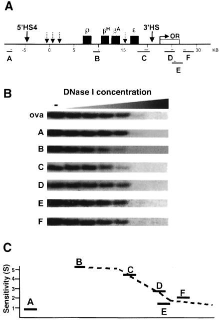
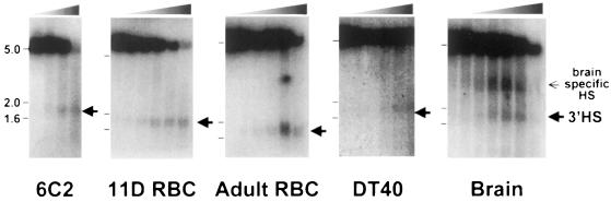
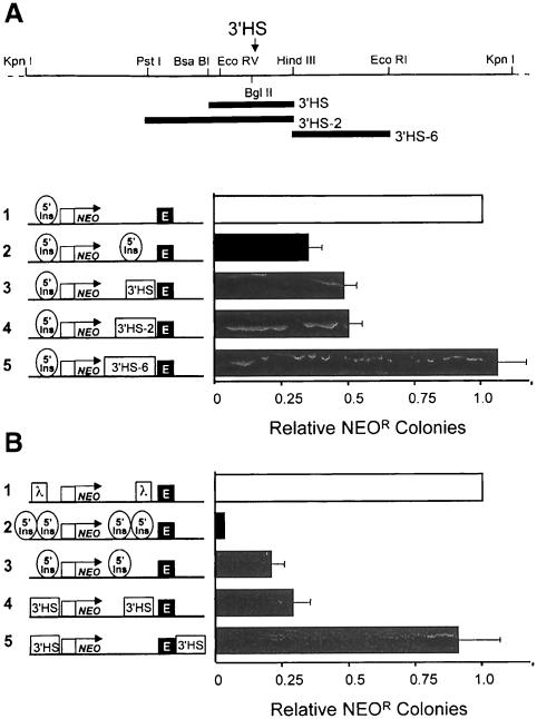
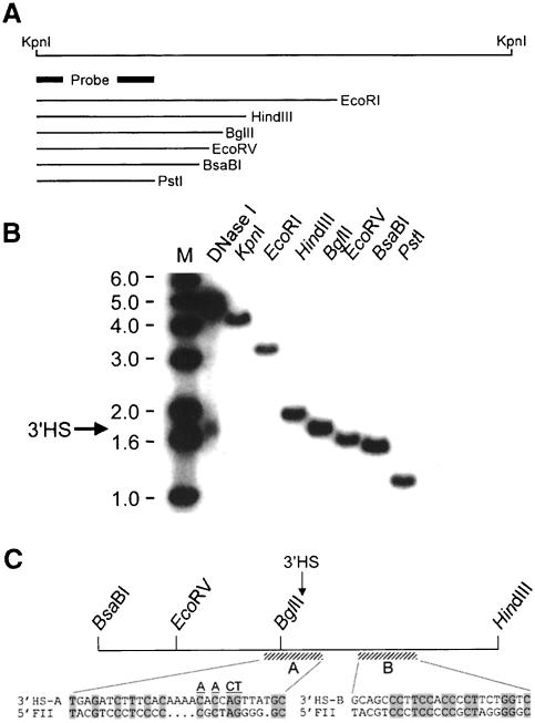
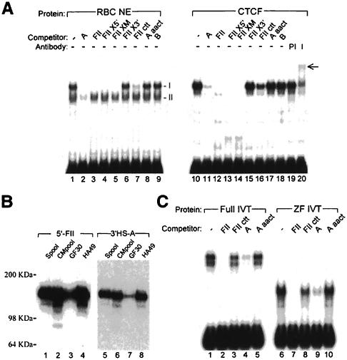
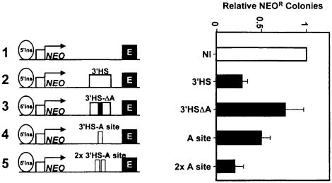
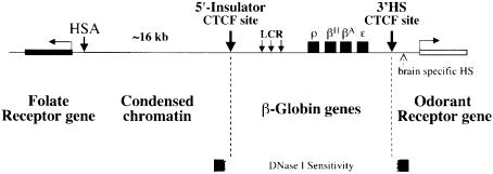
We show that the 3' boundary of the chicken beta-globin locus bears striking structural similarities to the 5' boundary. In erythroid cells a clear transition in DNase I sensitivity of chromatin at the 3' end of the locus is observed, the location of this transition is marked by a constitutive DNase I hypersensitive site (HS), and DNA spanning this site has the enhancer-blocking capacity of an insulator. This HS contains a binding site for the transcription factor CTCF. As in the case of the 5' insulator, the CTCF site is both necessary and sufficient for the enhancer-blocking activity of the 3' boundary. The position of this insulator is consistent with our proposal that it may function to maintain the distinct regulatory programs of the globin genes and their closely appended 3' neighbor, an odorant receptor gene. We conclude that both boundaries of the chicken beta-globin domain are capable of playing functionally similar roles and that the same protein is a necessary component of the molecular mechanism through which these boundaries are defined.


Figures

None
Fig. 1. A transition in DNase I sensitivity defines the 3′ boundary of the chicken β-globin domain. The 3′ boundary of generalized DNase I sensitivity is located between regions C and D. (A) Structure of the chicken β-globin locus is shown on the top. The four globin genes are indicated with black boxes. Selected HSs are shown by arrows. An open reading frame encoding the chicken odorant receptor protein (COR3′β) described in this study (Bulger et al., 1999) is shown as a white box marked ‘OR’. 5′HS4, the most upstream constitutive HS, marks the 5′ chromatin boundary and functions as a chromatin insulator. Another constitutive HS, 3′HS, was identified in this study. Other erythroid-specific HSs are shown by arrows with dotted lines. Numbers immediately below the map are map units based on the arbitrary numbering system set (Villeponteau and Martinson, 1981). Restriction fragments A–F detected in DNase I sensitivity assays in (B) are shown below the map. Probes used are indicated as thin lines. A detailed description of DNA fragments A–F and probes is given in Materials and methods. (B) Generalized DNase I sensitivity of DNA fragments A–F visualized by Southern blot hybridization. Erythrocyte nuclei isolated from 11-day-old chick embryos were treated with increasing amounts of DNase I (from right to left lanes, 0, 0.2, 0.4, 0.6, 1.0, 2.0 and 5.0 U/ml). Restriction fragments A–F were detected by Southern blot hybridizations. Relative sensitivities to DNase I correspond to the extent of loss of signal intensities of each band. DNA fragments B and C are relatively sensitive to DNase I, while fragments D–F are resistant to DNase I. A DNA fragment derived from the ovalbumin gene, which is transcriptionally inactive in this cell type, and fragment A, which is located farther upstream of the 5′ chromatin boundary, were used as DNase I resistant controls. (C) Quantitative representation of the DNase I sensitivities along the β-globin locus. Normalized DNase I sensitivity values (S) were calculated from the following equation (Pikaart et al., 1992) and plotted on a graph: S = log (GD/GU)/log (OD/OU) × T, where G and O are β-globin and ovalbumin band intensities for the undigested (U) or digested (D) samples and T is the size ratio of the ovalbumin to globin fragments. DNase I sensitivity drops significantly between C and D.
None
Fig. 2. A constitutive hypersensitive site (3′HS) within the 3′ chromatin boundary. Nuclei from various types of chicken cells were treated with increasing amounts of DNase I (from left to right lanes: 0, 0.06, 0.1, 0.2, 0.4 and 0.6 U/ml for RBCs and brain nuclei; 0, 8.0, 10, 20, 40 and 60 U/ml for DT40 and 6C2 cell nuclei). Genomic DNA was extracted and digested with KpnI. In addition to a parental fragment, a hypersensitive site, 3′HS, was detected as marked by arrowheads, in all cell types tested. Cells tested are a chicken erythroid precursor derived cell line (6C2), erythroid cells from either 11-day-old embryo (11D RBC) or adult blood (Adult RBC), brain from 11-day-old embryos (Brain) and a lymphoma-cell derived cell line (DT40).
None
Fig. 3. Directional enhancer-blocking activity of the 3′HS. (A) The human erythroleukemic cell line K562 was stably transfected with the constructs shown on the left. Each construct has the neomycin resistance gene (NEO) driven by a human βA-globin promoter with mouse β-globin HS2 as an enhancer. The DNA fragments 3′HS and 3′HS-2 include the DNase I HS 3′HS. 3′HS-6 does not contain the HS. For each construct, the 1.2 kb chromatin insulator fragment (5′Ins) including the 5′HS4 was placed upstream of the promoter in order to block influence from regulatory elements at the site of integration. The level of expression of each construct was measured as the number of neomycin-resistant colonies. Colony numbers obtained from construct 1, which does not have a DNA fragment between the promoter and the enhancer, were set at 100. Relative numbers of neomycin-resistant colonies are shown in the bar graph. We present the mean of five independent experiments. Enhancer-blocking activity resides in a DNA fragment containing the 3′HS. (B) Enhancer-blocking assays were performed using constructs shown to the left. In construct 1, a 2.3 kb fragment of λ DNA was inserted, as a spacer control, between the enhancer and the reporter. Thick bars show means of at least four independent experiments.
None
Fig. 4. Sequences homologous to the 5′ insulator element of the chicken β-globin locus are found at the site of the 3′HS. The position of the 3′HS was measured by the indirect end-labeling method and the strategy is shown in (A). Nuclei from 11-day-old chick embryos were treated with 0.4 U/ml DNase I, from which genomic DNA was extracted and digested with KpnI. In (B), the position of the 3′HS (arrow) was compared with the migration of genomic fragments of known length. The 3′HS hypersensitive fragment co-migrates with a fragment derived from BglII digestion. (C) Sequences homologous to FII from 5′HS4 (Chung et al., 1997; Bell et al., 1999) are found at or close to the sites of 3′HS, 3′HS-A and 3′HS-B, respectively. Alignment of the sequences 3′HS-A and 3′HS-B with the sequences of the 5′FII is shown. Conserved bases are shaded. Bases altered to generate a mutant site are underlined.
None
Fig. 5. Sequences derived from the 3′HS are bound specifically by CTCF. (A) Gel retardation analysis of complexes formed with the 3′HS-A sequence. Duplex 100mer oligonucleotides containing the 3′HS-A site were incubated with adult chicken RBC nuclear extracts (lanes 1–9) or partially purified CTCF (lanes 10–20; Bell et al., 1999). Two complexes observed following incubation with nuclear extracts are labeled I and II. Complexes were competed with 50-fold excesses of unlabeled competitor oligonucleotides containing either the 3′HS-A (lanes 2 and 11), 5′FII (lanes 3 and 12), FIIX5′-mutant (lanes 4 and 13), FIIXM-mutant (lanes 5 and 14), FIIX3′-mutant (lanes 6 and 15), FII ctt-mutant (lanes 7 and 16; Bell et al., 1999), 3′HS-A aact-mutant (lanes 8 and 17) or 3′HS-B (lanes 9 and 18) sites. Pre-immune antibodies (lane 19) or anti-CTCF antibodies (lane 20) were added to the binding reactions, and supershifted material is indicated by the arrow at the right. (B) Southwestern analysis of oligonucleotide duplexes binding to immobilized fractions of purified CTCF. Fractions of increased CTCF purity were separated by polyacrylamide gel electrophoresis and immobilized on PVDF membrane. The positions of molecular weight markers are indicated. A single gel blot was cut in two and probed with 32P-labeled oligonucleotide duplexes containing the 5′FII (lanes 1–4) and 3′HS-A (lanes 5–8) sites. Following washing, probed blots were exposed to autoradiographic film for 3 min and 2 h, respectively. (C) Gel retardation analysis of complexes formed between the 3′HS sequence and in vitro translated CTCF proteins. Duplex 100mer oligonucleotides containing the 3′HS-A site were incubated with in vitro translated proteins representing the full-length (lanes 1–5) or the zinc finger domain (lanes 6–10) of CTCF. Complexes were competed with 50-fold excesses of unlabeled competitor as indicated.
None
Fig. 6. The CTCF binding site found in the 3′HS is sufficient and necessary for enhancer-blocking activity. The enhancer-blocking activities of the 3′HS-A variants indicated were measured in the colony assay. In construct 3, a 56 bp fragment spanning the A-site has been deleted from the 400 bp 3′HS fragment and inserted into construct 1. One (construct 4) and two (construct 5) copies of the 3′HS-A site alone confer enhancer-blocking activity.
None
Fig. 7. Summary of the chromatin structure at the boundaries of the chicken β-globin locus in erythroid cells. A schematic view of the region extending from the chicken β-globin locus is shown. CTCF binds to both the 5′ and 3′ chromatin boundaries (Bell et al., 1999; this study), and may be involved in shielding the β-globin locus from regulatory effects from neighboring genes: the upstream folate receptor gene (Prioleau et al., 1999) and the downstream odorant receptor gene (Bulger et al., 1999; this study), respectively.

References

    1. Abe K., Kusakabe,Y., Tanemura,K., Emori,Y. and Arai,S. (1993) Multiple genes for G protein-coupled receptors and their expression in lingual epithelia. FEBS Lett., 316, 253–256. - PubMed
    1. Arnold R., Burcin,M., Kaiser,B., Muller,M. and Renkawitz,R. (1996) DNA bending by the silencer protein NeP1 is modulated by TR and RXR. Nucleic Acids Res., 24, 2640–2647. - PMC - PubMed
    1. Barton M.C., Hoekstra,M.F. and Emerson,B.M. (1990) Site-directed, recombination-mediated mutagenesis of a complex gene locus. Nucleic Acids Res., 18, 7349–7355. - PMC - PubMed
    1. Bell A.C. and Felsenfeld,G. (1999) Stopped at the border: boundaries and insulators. Curr. Opin. Genet. Dev., 9, 191–198. - PubMed
    1. Bell A.C., West,A.G. and Felsenfeld,G. (1999) The protein CTCF is required for the enhancer-blocking activity of vertebrate insulators. Cell, 98, 387–396. - PubMed

Publication types