. 2000 May 15;19(10):2351-61.
doi: 10.1093/emboj/19.10.2351.

The 2 A crystal structure of leucyl-tRNA synthetase and its complex with a leucyl-adenylate analogue

Affiliations

The 2 A crystal structure of leucyl-tRNA synthetase and its complex with a leucyl-adenylate analogue

S Cusack et al. EMBO J. .

Abstract

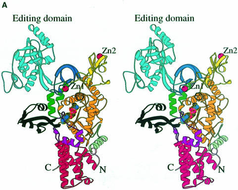
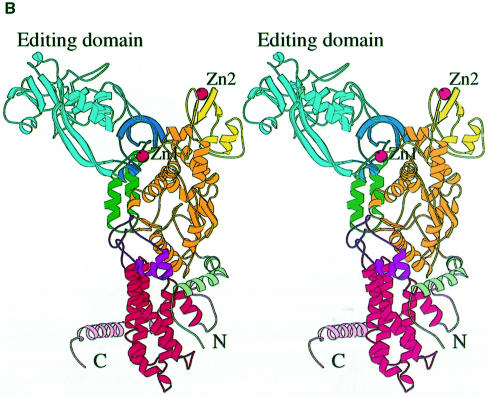
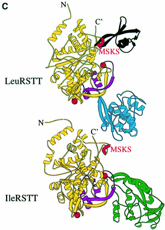
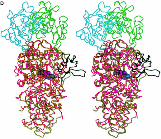
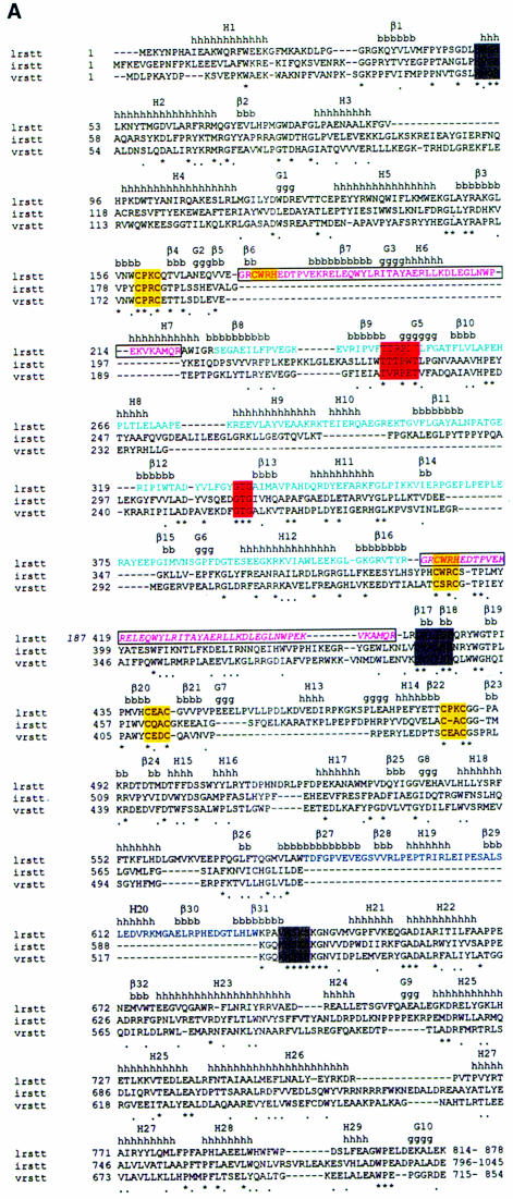
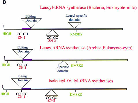
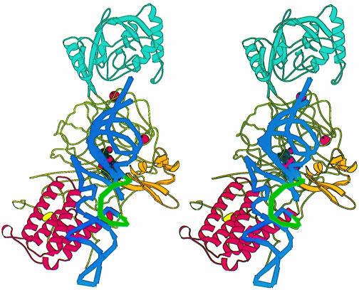
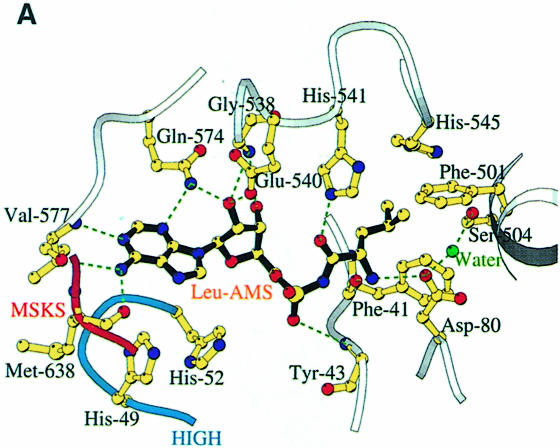
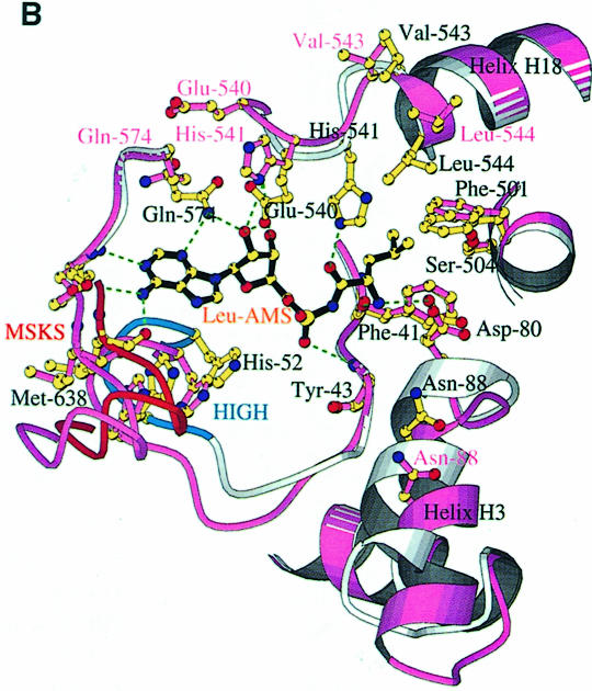
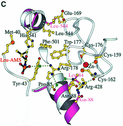
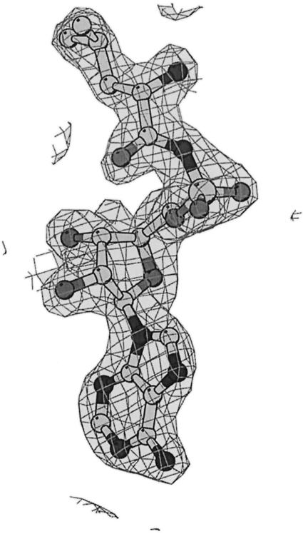
Leucyl-, isoleucyl- and valyl-tRNA synthetases are closely related large monomeric class I synthetases. Each contains a homologous insertion domain of approximately 200 residues, which is thought to permit them to hydrolyse ('edit') cognate tRNA that has been mischarged with a chemically similar but non-cognate amino acid. We describe the first crystal structure of a leucyl-tRNA synthetase, from the hyperthermophile Thermus thermophilus, at 2.0 A resolution. The overall architecture is similar to that of isoleucyl-tRNA synthetase, except that the putative editing domain is inserted at a different position in the primary structure. This feature is unique to prokaryote-like leucyl-tRNA synthetases, as is the presence of a novel additional flexibly inserted domain. Comparison of native enzyme and complexes with leucine and a leucyl- adenylate analogue shows that binding of the adenosine moiety of leucyl-adenylate causes significant conformational changes in the active site required for amino acid activation and tight binding of the adenylate. These changes are propagated to more distant regions of the enzyme, leading to a significantly more ordered structure ready for the subsequent aminoacylation and/or editing steps.


Figures

None
Fig. 1. (A) Stereo ribbon diagram of the structure of the complex between T.thermophilus leucyl-tRNA synthetase and a leucyl-adenylate analogue. The domains are coloured as follows: N-terminal extension (pale green), catalytic domain (Rossmann-fold, orange), ZN-1 domain (blue), helical hairpin insertion (green), editing domain (cyan), ZN-2 domain (yellow), leucyl-specific insertion domain (black), connecting module (purple), anti-codon binding domain (red) and C-terminal extension (pink). The zinc atoms are shown as red balls and the leucyl-adenylate analogue as a space-filling model in the centre. The N- and C-termini are marked. (B) Stereo ribbon diagram of the structure of the T.thermophilus isoleucyl-tRNA synthetase. Colouring as in (A). Note that in IleRS the ZN-1 domain is split by the inserted editing domain [see (C) and Figure 2]. (C) Comparison of point of insertion of extra domains in T.thermophilus leucyl- (top) and isoleucyl- (bottom) tRNA synthetases. For clarity, the enzymes are C-terminally truncated just before the anti-codon binding domain (C′). The purple polypeptide segment is homologous in each enzyme and contains the second half of the ZN-1 binding site [see also (A)]. In leucyl-tRNA synthetase, it precedes the editing domain (cyan), whereas in isoleucyl-tRNA synthetase it follows the editing domain (green). The disposition of the leucyl-specific insertion domain is shown in black. It is inserted just before the KMSKS loop (red segment in both enzymes). (D) Stereo view of the superposition of Cα traces of LeuRSTT and IleRSTT showing different points of insertion of the editing domain and the near 180° difference in rotational orientation. LeuRSTT is red with editing domain cyan, leucine-specific domain black and LeuAMS in the active site. IleRSTT is yellow with editing domain green. Considering the red and yellow traces, 437 Cα positions superpose within a cut-off of 3.8 Å with 115 identities and root-mean-square deviation (r.m.s.d.) of 1.99 Å. Superposing the two editing domains, the equivalent figures are 129 Cα positions with 37 identities and r.m.s.d. of 1.36 Å. In all, 69.5% of the 814 ordered residues in LeuRSTT can be superposed within 3.8 Å of equivalent residues in IleRSTT with 27% of these residues being identical.
None
Fig. 2. (A) Primary sequence alignment of T.thermophilus leucyl-, isoleucyl- and valyl-tRNA synthetases. The alignment of LeuRSTT and IleRSTT is based on superposition of three-dimensional structures (see the legend to Figure 1D). An asterisk indicates amino acid identity and a stop indicates amino acid similarity. The secondary structure elements as defined by DSSP (Kabsch and Sander, 1983) are indicated for LeuRSTT (h, α-helix; g, 310 helix; β, β-strand). The editing domain of LeuRSTT is shown with cyan letters, the leucyl-specific domain with blue letters. The Class 1a conserved motifs, HIGH, DWLISR (Carter, 1993) and KMSKS, are boxed in grey. The conserved threonine-rich and GTG motifs of the editing domain are boxed in red. Zinc binding motifs are boxed in yellow. The boxed, pink region in LeuRS is shown twice: first in its actual position in the primary sequence (residues 174–211) and secondly (in italics) displaced to after the editing domain where it aligns with corresponding sequences in IleRS and ValRS. This peptide is also coloured purple in Figure 1C. Sequences are C-terminally truncated after the end of the visible region in the LeuRSTT structure. (B) Schematic diagram of the domain structure of leucyl-, isoleucyl- and valyl-tRNA synthetases, based on extensive sequence alignments. The positions of the two catalytically important class I motifs, HIGH and MSKS, are shown. The purple segment is homologous in each enzyme. Top: domain structure of T.thermophilus leucyl-tRNA synthetase, representative of eubacterial and mitochondrial leucyl-tRNA synthetases. Although the ZN-1 and ZN-2 domains are always conserved, the zinc ligands are not, so that there can be both zinc atoms (e.g. T.thermophilus, Helicobacter pylori), no zinc atoms (e.g. Borrelia burgdorferi, Mycoplasma), Zn-1 only (e.g. Haemophilus influenzae, E.coli) or Zn-2 only (human mitochondrial, Rickettsia prowazekii). A similar variability of zinc content is found in methionyl-tRNA synthetase (Mechulam, 1999). The specific insertion domain (black) ranges in size from 30 to 60 residues. Interestingly, the putative Caenorhabditis elegans mitochondrial leucyl-tRNA synthetase has a severely truncated and probably inactive editing domain. Middle: predicted domain structure of archae and eukaryotic cytoplasmic leucyl-tRNA synthetases. The editing domain splits the ZN-1 domain. Putative zinc ligands are present in the ZN-1 domain generally for archae enzymes, but not for eukaryotic cytoplasmic enzymes. Sequence alignments suggest that there is no ZN-2 domain, which normally precedes one of the leucine binding motifs (containing Phe501 and Ser504 in LeuRSTT), although there seems to be a short specific insertion after this leucine binding motif. Bottom: domain structure of T.thermophilus isoleucyl-tRNA synthetase (Nureki et al., 1998), representative of all isoleucyl- and valyl-tRNA synthetases.
None
Fig. 3. Stereo diagram of a model of a putative LeuRSTT-tRNAleu editing complex obtained by superimposition of LeuRSTT on the IleRSSA-tRNAile editing complex (Silvian et al., 1999). The model tRNA used here is that of tRNAtyr with a long variable arm (S.Cusack, A.Yaremchuk and M.Tukalo, unpublished results). Its position has been manually adjusted to avoid steric clashes with the enzyme, notably with the ZN-1 domain, which is much closer to the active site in LeuRSTT than in the IleRSSA-tRNAile complex. The anticodon binding domain is in red, the editing domain in cyan. The model shows that the leucyl-specific domain (orange) could interact with the base of the acceptor stem and core of the tRNA. The long variable arm of the tRNA (green) points away from the enzyme and towards the viewer. This is consistent with it not being an important identity element, although it is possible that the disordered C-terminal domain of the enzyme makes some contact with this part of the tRNA. Given the flexibility of the various modules of the enzyme and of the tRNA itself, the aim of this figure is to give the general disposition of the various elements rather than to be an accurate model.
None
Fig. 4. (A) The LeuAMS binding site showing the major interacting residues. Hydrogen bonds are shown as dashed green lines and a tightly bound water as a green sphere. The catalytically essential class 1 motifs H49IGH and M638SKS are shown in cyan and red, respectively. The side chain of Tyr43 is omitted for clarity, but is visible in (C). (B) The conformational changes associated with LeuAMS binding. The view is the same as in (A). The pink ribbon diagram, pink side chains and pink labels correspond to the apo-structure (mercury derivative) and the grey ribbon and yellow side chains belong to the LeuAMS-bound structure. Upon binding of the adenosine moiety, the HIGH and MSKS loops towards the active centre, Gln574 and Glu540 move to bind the ribose tightly, and helices H18 and H3 refold to permit packing of the ZN-1 domain close to the active site [see the text and (C)]. A sulfate ion (not shown) is bound to His49 and His52 in the apo-structure, but not in the LeuAMS-bound structure. (C) Proximity of Arg178 to the active center in the LeuAMS complex. Colouring as in (B) with water molecules as green spheres and the Zn-1 atom as a red sphere. One of the zinc ligands (His179) and the adenosine moiety of the LeuAMS are omitted for clarity. The positions of Leu544 and Leu84 sterically prevent the packing of the ZN-1 domain close to the active site in the apo-structure.
None
Fig. 5. Final electron density at 2.0 Å resolution for LeuAMS in the active site of LeuRSTT contoured at 2 σ.

References

    1. Asahara H., Himeno,H., Tamura,K., Hasegawa,T., Watanabe,K. and Shimizu,M. (1993) Recognition nucleotides of Escherichia coli tRNALeu and its elements facilitating discrimination from tRNASer and tRNATyr. J. Mol. Biol., 231, 219–229. - PubMed
    1. Brünger A.T. et al. (1998) Crystallographic and NMR system: a new software suite for macromolecular structure determination. Acta Crystallogr. D, 54, 905–921. - PubMed
    1. Carter C.W. Jr (1993) Cognition, mechanism and evolutionary relationships in aminoacyl-tRNA synthetases. Annu. Rev. Biochem., 62, 715–748. - PubMed
    1. Cavarelli J., Delagoutte,B., Eriani,G., Gangloff,J. and Moras,D. (1998) l-arginine recognition by yeast arginyl-tRNA synthetase. EMBO J., 17, 5438–5448. - PMC - PubMed
    1. Collaborative Computational Project Number 4 (1994) The CCP4 suite: programs for protein crystallography. Acta Crystallogr. D, 50, 760–763. - PubMed

MeSH terms