. 2000 May 15;525 Pt 1(Pt 1):135-42.
doi: 10.1111/j.1469-7793.2000.00135.x.

An oxygen-, acid- and anaesthetic-sensitive TASK-like background potassium channel in rat arterial chemoreceptor cells

Affiliations

An oxygen-, acid- and anaesthetic-sensitive TASK-like background potassium channel in rat arterial chemoreceptor cells

K J Buckler et al. J Physiol. .

Abstract

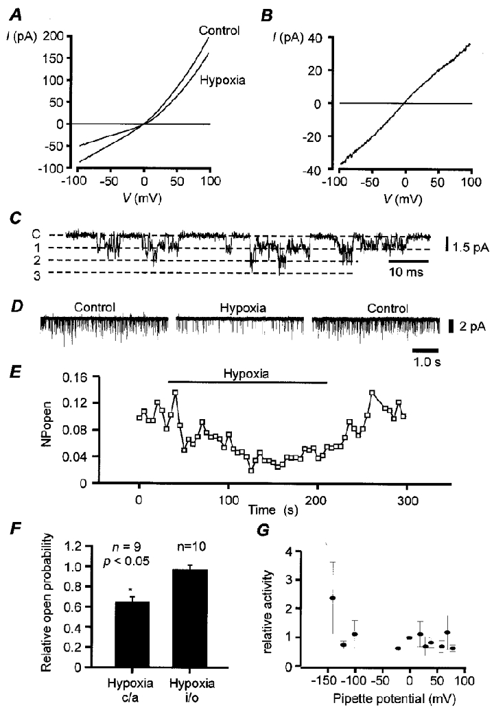
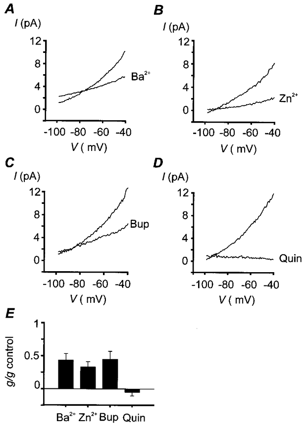
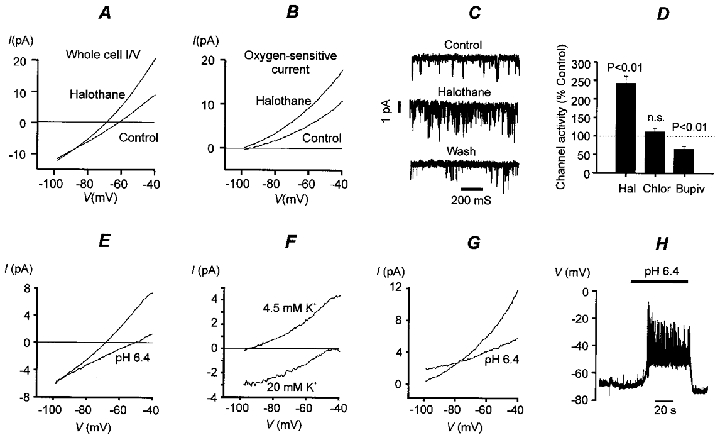
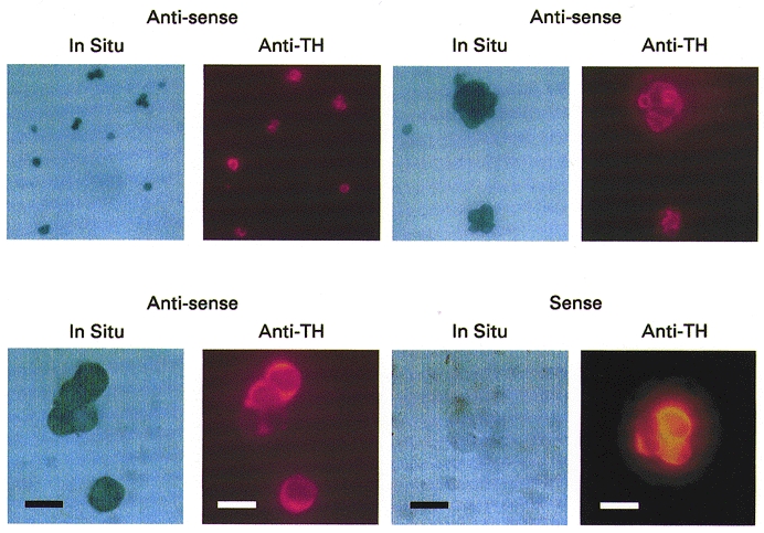
The biophysical and pharmacological properties of an oxygen-sensitive background K+ current in rat carotid body type-I cells were investigated and compared with those of recently cloned two pore domain K+ channels. Under symmetrical K+ conditions the oxygen-sensitive whole cell K+ current had a linear dependence on voltage indicating a lack of intrinsic voltage sensitivity. Single channel recordings identified a K+ channel, open at resting membrane potentials, that was inhibited by hypoxia. This channel had a single channel conductance of 14 pS, flickery kinetics and showed little voltage sensitivity except at extreme positive potentials. Oxygen-sensitive current was inhibited by 10 mM barium (57% inhibition), 200 microM zinc (53% inhibition), 200 microM bupivacaine (55% inhibition) and 1 mM quinidine (105 % inhibition). The general anaesthetic halothane (1.5%) increased the oxygen-sensitive K+ current (by 176%). Halothane (3 mM) also stimulated single channel activity in inside-out patches (by 240%). Chloroform had no effect on background K+ channel activity. Acidosis (pH 6.4) inhibited the oxygen-sensitive background K+ current (by 56%) and depolarised type-I cells. The pharmacological and biophysical properties of the background K+ channel are, therefore, analogous to those of the cloned channel TASK-1. Using in situ hybridisation TASK-1 mRNA was found to be expressed in type-I cells. We conclude that the oxygen- and acid-sensitive background K+ channel of carotid body type-I cells is likely to be an endogenous TASK-1-like channel.


Figures

Figure 1
Figure 1. Biophysical properties of background K+ channels
A, mean whole cell current-voltage relationship (I–V) for type-I cells bathed in 140 mM K+,Ca2+-free Tyrode solution containing 2.5 mM Ni2+, 10 mM TEA and 5 mM 4-AP, under normoxic and hypoxic conditions (n = 10). Hypoxia was induced by gassing saline solutions with 5 % CO2-95 % N2. Hypoxia reduced membrane conductance (over voltage range -70 to -60 mV) from 0.84 ± 0.12 nS control to 0.49 ± 0.09 nS hypoxia (n = 10; P < 0.001). Mean currents at -50 mV were 46.4 ± 0.6 pA control, and 26.4 ± 3.0 pA hypoxia. B, mean oxygen-sensitive current determined by subtraction of I–V obtained under hypoxic conditions from that obtained under normoxic conditions (from data in A). C, single channel recording from a cell-attached patch. Cell bathed in standard Tyrode solution, pipette solution contained 10 mM TEA, pipette potential 0 mV. D and E, hypoxia reversibly reduces channel activity in cell-attached patch. Cells were bathed in 100 mM potassium bicarbonate-buffered Tyrode solution (K+ substituted for Na+). Pipette potential was 70 mV (membrane potential approx. -70 to -80 mV). F, effects of hypoxia on single channel activity in cell-attached (c/a) patches and inside-out (i/o) patches. Hypoxia significantly reduced channel activity in cell-attached but not inside-out patches (*). Inside-out patches were clamped at -100 mV. Data expressed as fraction of control NPopen. G, channel activity, relative to that observed at the resting potential (0 mV pipette potential), as a function of pipette potential for cell-attached patch (n = 5). Pipette solution contained 10 mM TEA and 5 mM 4-AP. Cells bathed in standard bicarbonate Tyrode solution.
Figure 2
Figure 2. Pharmacology of background K+ channels
Oxygen-sensitive currents were determined by subtraction of currents obtained under hypoxic conditions from those obtained under normoxic conditions, both in the absence and in the presence of inhibitor. Experiments were performed in normal, physiological, bicarbonate-buffered saline, [K+]o= 4.5 mM; note that under these conditions the oxygen-sensitive current shows weak outward rectification. Data are mean I–V relationships for a number of cells. The degree of inhibition was determined by calculation of membrane conductance in the region -60 to -70 mV. A, effects of 10 mM Ba2+. Mean conductance: 161 ± 30 pS control, 61 ± 15 pS Ba2+, n = 9, P < 0.01 (Student's paired t test). Mean oxygen-sensitive current at -50 mV: 7.8 ± 1.0 pA control, 4.9 ± 0.8 pA Ba2+. B, effects of 200 μM Zn2+. Mean conductance: 160 ± 28 pS control, 47 ± 13 pS Zn2+, n = 6, P < 0.01. Mean oxygen-sensitive current at -50 mV: 6.1 ± 1.0 pA control, 2.0 ± 0.7 pA Zn2+. C, effects of 200 μM bupivacaine (Bup). Mean conductance: 218 ± 33 pS control, 93 ± 22 pS bupivacaine, n = 6, P < 0.005. Mean oxygen-sensitive current at -50 mV: 8.7 ± 1.6 pA control, 5.2 ± 0.6 pA bupivacaine. D, effects of 1 mM quinidine (Quin). Mean conductance: 219 ± 37 pS control, -11 ± 11 pS quinidine, n = 5, P < 0.005. Mean oxygen-sensitive current at -50 mV: 9.7 ± 1.5 pA control, 0.6 ± 0.1 pA quinidine. E, summary of effects of K+ channel inhibitors on oxygen-sensitive membrane conductance. Mean percentage reduction in conductance: Ba2+, 57 ± 10 %; Zn2+, 67 ± 8 %; bupivacaine, 55 ± 12 %; quinidine, 105 ± 5 %.
Figure 3
Figure 3. Effects of anaesthetics and acidosis on background K+ channels
A, effect of halothane on whole cell conductance. Mean I–V plot from 9 cells. Mean conductance: 350 ± 36 pS control, 584 ± 68 pS halothane, n = 9, P < 0.001. Mean whole cell current at -50 mV: 4.9 ± 0.9 pA control, 12.6 ± 1.9 pA halothane. B, effects of halothane on mean oxygen-sensitive currents (determined by subtraction, control – hypoxia). Mean oxygen-sensitive conductance: 183 ± 33 pS control, 323 ± 64 pS halothane, n = 9, P < 0.02. Mean oxygen-sensitive current at -50 mV: 7.6 ± 1.4 pA control, 13.1 ± 2.6 pA halothane. Halothane was applied by gassing saline solutions with 1.5 % halothane-5 % CO2-20 % O2-73.5 % N2 (control) or 1.5 % halothane-5 % CO2-93.5 % N2 (hypoxia). C, 3 mM halothane applied intracellularly to inside-out patches increases channel activity. Membrane potential, -70 mV. D, comparison of effects of local and general anaesthetics on single channel activity in inside-out patches. Hal, 3 mM halothane; membrane potential -70 mV, n = 10. Chlor, 3 mM chloroform; membrane potential -70 mV, n = 7. Bupiv, 500 μM bupivacaine; membrane potential -100 mV, n = 8. E, effect of isocapnic acidosis (pH 6.4) on whole cell membrane conductance (mean I–V from 5 cells). Cells were exposed to acid solution for 20 s periods every 1–2 min. Note that acidosis reduces membrane conductance (control, 270 ± 22 pS; acidosis, 131 ± 19 pS; n = 5, P < 0.001) and shifts the zero current potential in a positive (depolarising) direction. Mean current at -50 mV: 4.6 ± 0.6 pA control, -0.6 ± 0.7 pA pH 6.4. F, mean acid-sensitive currents (control – acidosis; n = 6) determined at two different levels of extracellular K+ (acid stimuli applied for 1–2 min). Note leftward/downward shift of I–V in high K+ and depolarising shift in reversal potential. Mean acid-sensitive current at -50 mV: 4.6 ± 0.6 pA in 4.5 mM [K+]o, -0.4 ± 0.5 pA in 20 mM [K+]o. G, effects of acidosis (2 min) on oxygen-sensitive current. Acidosis reduced the oxygen-sensitive conductance from 203 ± 32 pS to 88 ± 21 pS (measured over voltage range -70 to -60 mV). Mean oxygen-sensitive current at -50 mV: 8.3 ± 1.3 pA control, 4.8 ± 1.0 pA pH 6.4. H, recording of membrane potential in a type-I cell during exposure to isocapnic acidosis. Experiments depicted in parts A, B, E, F, G and H were performed in a normal, physiological, bicarbonate-buffered saline, [K+]o= 4.5 mM. Inside-out patch recordings shown in C and D were performed in a Hepes-buffered saline with high [K+]o (see Methods).
Figure 4
Figure 4. In situ hybridisation for TASK-1
Staining for digoxigenin-labelled RNA probes with anti-digoxigenin-alkaline phosphatase (panels labelled In Situ). The same cells were also stained with anti-tyrosine hydroxylase (shown in adjacent panels labelled Anti-TH). Three examples of in situ hybridisation with anti-sense probes for TASK-1 are shown at increasing magnification (top row and bottom left), and one example of in situ hybridisation with sense probes (bottom right). Scale bar = 10 μm (bottom row only). Note that only anti-sense probes produced staining of cells, and that both type-I cells (positive for tyrosine hydroxylase) and possibly non-type-I cells (weak or no anti-tyrosine hydroxylase staining) express TASK-1 mRNA.

References

    1. Bargmann CI. Neurobiology of the Caenorhabditis elegans genome. Science. 1998;282:2028–2033. - PubMed
    1. Buckler KJ. A novel oxygen-sensitive potassium current in rat carotid body type I cells. The Journal of Physiology. 1997;498:649–662. - PMC - PubMed
    1. Buckler KJ, Vaughan-Jones RD. Effects of acidic stimuli on intracellular calcium in isolated type-I cells of the neonatal rat carotid body. Pflügers Archiv. 1993;425:22–27. - PubMed
    1. Buckler KJ, Vaughan-Jones RD. Effects of hypoxia on membrane potential and intracellular calcium in rat neonatal carotid body type-I cells. The Journal of Physiology. 1994a;476:423–428. - PMC - PubMed
    1. Buckler KJ, Vaughan-Jones RD. Effects of hypercapnia on membrane potential and intracellular calcium in rat carotid body type-I cells. The Journal of Physiology. 1994b;478:157–171. - PMC - PubMed

Publication types

MeSH terms

LinkOut - more resources