. 2000 May 15;149(4):767-74.
doi: 10.1083/jcb.149.4.767.

Structural transitions at microtubule ends correlate with their dynamic properties in Xenopus egg extracts

Affiliations

Structural transitions at microtubule ends correlate with their dynamic properties in Xenopus egg extracts

I Arnal et al. J Cell Biol. .

Abstract

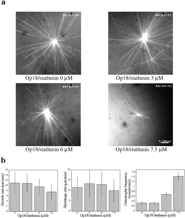
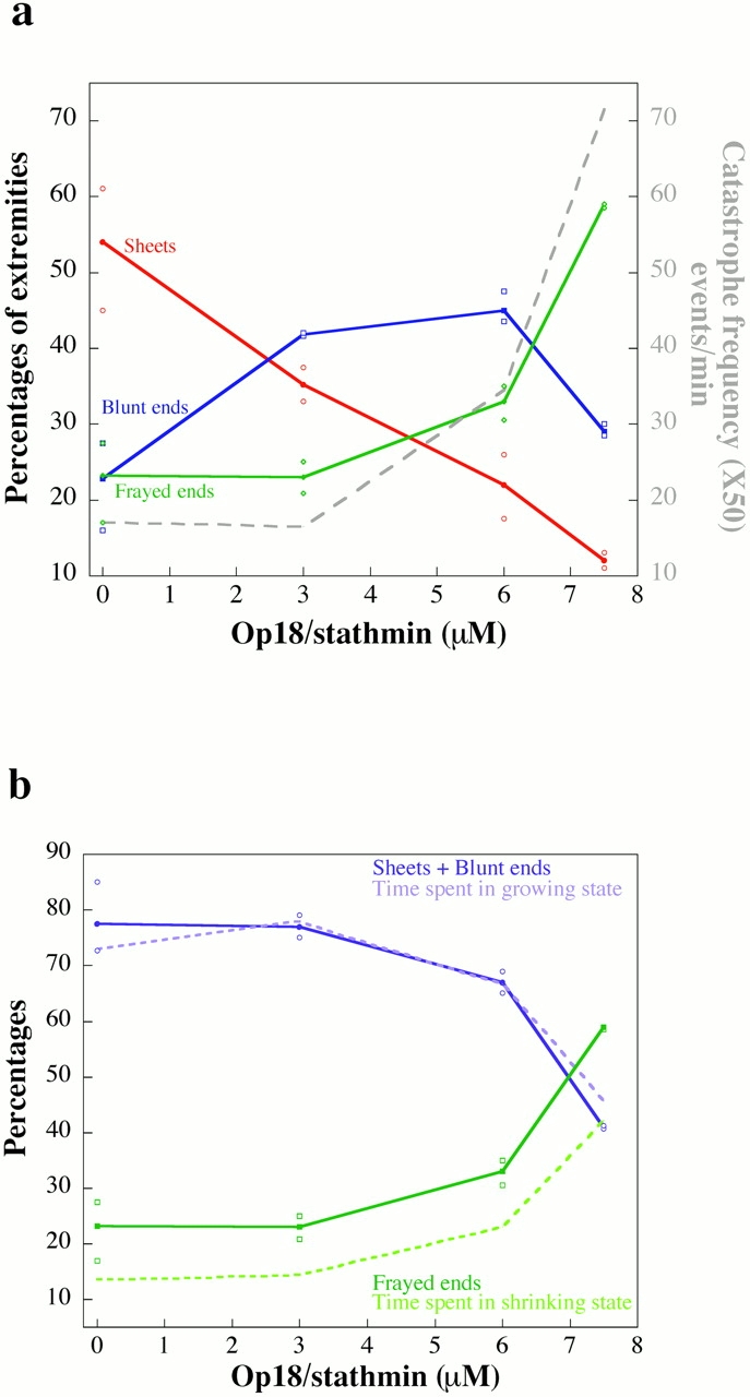
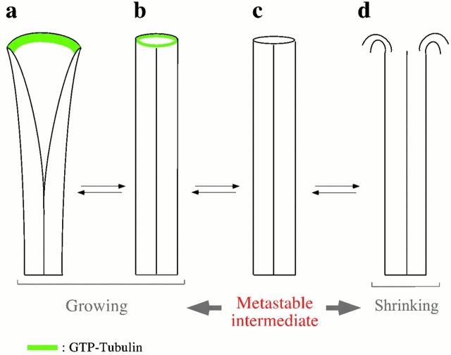
Microtubules are dynamically unstable polymers that interconvert stochastically between growing and shrinking states by the addition and loss of subunits from their ends. However, there is little experimental data on the relationship between microtubule end structure and the regulation of dynamic instability. To investigate this relationship, we have modulated dynamic instability in Xenopus egg extracts by adding a catastrophe-promoting factor, Op18/stathmin. Using electron cryomicroscopy, we find that microtubules in cytoplasmic extracts grow by the extension of a two- dimensional sheet of protofilaments, which later closes into a tube. Increasing the catastrophe frequency by the addition of Op18/stathmin decreases both the length and frequency of the occurrence of sheets and increases the number of frayed ends. Interestingly, we also find that more dynamic populations contain more blunt ends, suggesting that these are a metastable intermediate between shrinking and growing microtubules. Our results demonstrate for the first time that microtubule assembly in physiological conditions is a two-dimensional process, and they suggest that the two-dimensional sheets stabilize microtubules against catastrophes. We present a model in which the frequency of catastrophes is directly correlated with the structural state of microtubule ends.


Figures

Figure 1
Figure 1
(a) Vitreous ice–embedded microtubules observed in interphasic extracts supplemented with isolated centrosomes and incubated for 20 s at 25°C. E, extensions observed at microtubule ends; and C, centriole. (b and c) Vitreous ice–embedded microtubules observed in interphasic extracts in the presence of 7.5 μM recombinant Op18/stathmin. Addition of Op18/stathmin in this condition induces the disappearance of sheets at microtubule ends (black arrows). F, frayed end; and B, blunt end. Bar, 200 nm.
Figure 2
Figure 2
Detailed views of microtubule end structures in interphasic extracts. (a) Microtubule end with peeling protofilaments (frayed end). (b) Blunt end. (c–e) Extensions with variable lengths. Bar, 100 nm.
Figure 3
Figure 3
(a) Op18/stathmin shortens microtubules in interphasic extracts. Images were taken from videos at the same time point after the beginning of the recording in the presence of 0, 3, 6, and 7.5 μM of recombinant Op18/stathmin added in interphasic extracts. (b) Op18/stathmin increases the catastrophe frequency in interphasic extract. The growth rate, shrinkage rate, and catastrophe frequency were determined from the movies recorded over time in interphasic extract with a different amount of Op18/stathmin. The rescue frequency is not reported because the number of events recorded was too low in most of the conditions.
Figure 4
Figure 4
(a) Microtubule end structure in the presence of a different amount of recombinant Op18/stathmin added in interphasic extracts. Microtubule ends are represented as the percentage of the total number of extremities. Values are the mean of four (control) or two (3, 6, and 7.5 μM Op18/stathmin) experiments made in different extracts. For each condition, the mean value is represented by a closed symbol and the minimum and the maximum values by open symbols. (b) Microtubule end structure of growing and shrinking microtubules. The percentage of time spent in the growing state (light violet curve) as well as the combined proportion of sheets and blunt ends (dark violet curve) decrease with increased amount of recombinant Op18/stathmin added in interphasic extracts caused by the higher level of catastrophes. The percentage of time spent in the shrinking state (dark green curve) and the frayed end proportion (light green curve) follow the inverse change versus the Op18/stathmin concentration.
Figure 5
Figure 5
Structural model explaining dynamic instability and its possible relationship with the GTP cap model. (a) Microtubules with two-dimensional sheets at their end are in a stable growing state. GTP-tubulin (in green) contained in the sheet could explain part of its stabilization capacity. When the tube closes, a few subunits in a GTP state at the extremity would be sufficient to maintain the microtubule in a growing state. (b) As soon as the GTP is hydrolyzed (c), the microtubule will explode because of the weak lateral interaction between GDP-tubulin (d). Blunt ends (b and c) are unstable because the loss of the GTP-tubulin subunits triggers them in a shrinking state, whereas sheets are always stable.

References

    1. Amos L. The microtubule lattice20 years on. Trends Cell Biol. 1995;5:48–51. - PubMed
    1. Bayley P.M., Schilstra M.J., Martin S.R. Microtubule dynamic instabilitynumerical simulation of microtubule transition properties using a lateral cap model. J. Cell Sci. 1990;95:33–48. - PubMed
    1. Belmont L.D., Mitchison T.J. Identification of a protein that interacts with tubulin dimers and increases the catastrophe rate of microtubules. Cell. 1996;84:623–631. - PubMed
    1. Belmont L.D., Hyman A.A., Sawin K.E., Mitchison T.J. Real-time visualization of cell cycle-dependent changes in microtubule dynamics in cytoplasmic extracts. Cell. 1990;62:579–589. - PubMed
    1. Bornens M., Paintrand M., Berges J., Marty M.C., Karsenti E. Structural and chemical characterization of isolated centrosomes. Cell Motil. Cytoskeleton. 1987;8:238–249. - PubMed

Publication types