. 2000 May;105(10):1353-61.
doi: 10.1172/JCI8862.

Cubilin is an albumin binding protein important for renal tubular albumin reabsorption

Affiliations

Cubilin is an albumin binding protein important for renal tubular albumin reabsorption

H Birn et al. J Clin Invest. 2000 May.

Abstract

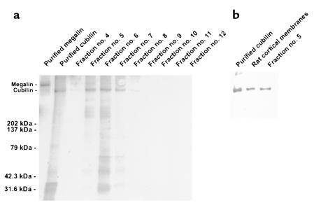
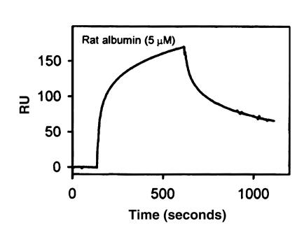
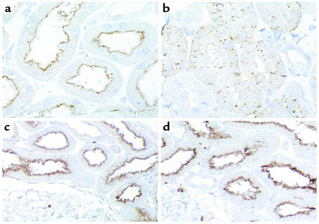
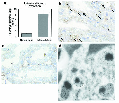
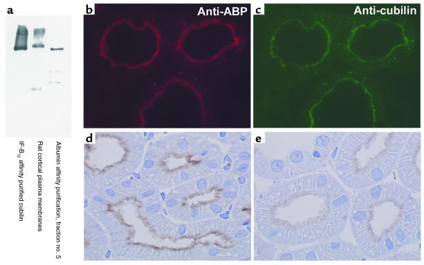
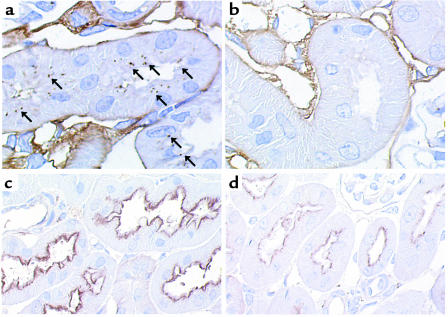
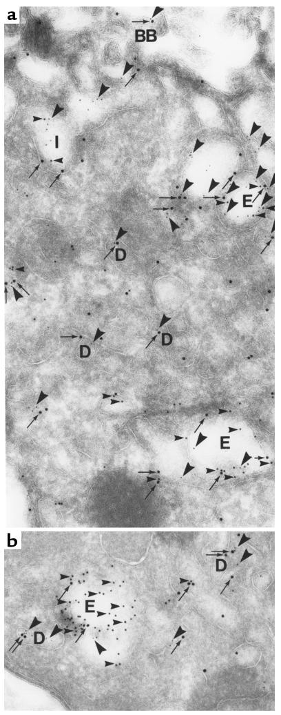
Using affinity chromatography and surface plasmon resonance analysis, we have identified cubilin, a 460-kDa receptor heavily expressed in kidney proximal tubule epithelial cells, as an albumin binding protein. Dogs with a functional defect in cubilin excrete large amounts of albumin in combination with virtually abolished proximal tubule reabsorption, showing the critical role for cubilin in the uptake of albumin by the proximal tubule. Also, by immunoblotting and immunocytochemistry we show that previously identified low-molecular-weight renal albumin binding proteins are fragments of cubilin. In addition, we find that mice lacking the endocytic receptor megalin show altered urinary excretion, and reduced tubular reabsorption, of albumin. Because cubilin has been shown to colocalize and interact with megalin, we propose a mechanism of albumin reabsorption mediated by both of these proteins. This process may prove important for understanding interstitial renal inflammation and fibrosis caused by proximal tubule uptake of an increased load of filtered albumin.


Figures

Figure 1
Figure 1
Albumin affinity purification of cubilin. (a) Eluted fractions were analyzed by SDS-PAGE and Coomassie stained. Lane 1: 0.1 μg of RAP affinity purified rabbit megalin; lane 2: IF-B12 affinity purified cubilin. Lanes 3–11: fractions 4–12 (12 μL/lane). The positions of standard molecular weight markers are indicated. A strong band corresponding to the size of cubilin is clearly identified in fractions 5–8. Faint low–molecular-weight bands are seen with both the IF-B12 purified and albumin purified cubilin. (b) The eluted protein was further identified as cubilin by immunoblotting using anti-rat cubilin antibodies.
Figure 2
Figure 2
SPR sensorgram of the binding of rat albumin to purified rat cubilin immobilized to a BIAcore sensor chip. The BIAevaluation software estimated Kd = 0.63 μM when fitting the curves to a one-site model. The on and off rates for binding of rat albumin were recorded by a flow of 5 μM albumin followed by flow of buffer alone. The SPR signal is displayed as the mass-equivalent RUs.
Figure 3
Figure 3
Localization of cubilin (a and b) and megalin (c and d) in kidney cortex of normal (a and c) and affected (b and d) dogs. Sections were incubated with anti-dog cubilin (1:4,000). In normal dogs, cubilin is localized in luminal plasma membranes of kidney proximal tubules (a), whereas the labeling in affected dogs is intracellular and vesicular with no detectable receptor in the luminal plasma membrane (b), suggesting retention in an intracellular compartment. No difference in the labeling intensity or in the distribution of megalin is observed between normal (c) and affected (d) dogs when evaluated by immunocytochemistry using a sheep anti-rat megalin (1:10,000). ×600 (a and b); ×350 (c and d).
Figure 4
Figure 4
Increased urinary excretion and decreased tubular reabsorption of albumin in affected dogs. (a) Dogs with a functional defect in cubilin expression (n = 6) reveals a significant, approximately sevenfold increase (P = 0.00003) in urinary albumin/creatinine excretion compared with normal control dogs (n = 6). (b and c) Immunohistochemistry using an anti-dog albumin antibody (1:100,000) to identify albumin in the kidney cortex of normal (b) and affected (c) dogs. In normal dogs (b), evidence of reabsorbed and endocytosed albumin in proximal tubules is clear (arrows), whereas no albumin can be identified inside the proximal tubules of affected dogs (c). In all dogs, a faint labeling was observed in basement membranes due to a basal deposition of albumin. ×600. Three affected and three normal dogs were examined using two different anti-albumin antibodies, all showing the same labeling pattern. (d) Electron microscopic immunocytochemistry using anti-dog albumin antibody (1:5,000) on a section of normal dog proximal tubule cells. Intense labeling of four lysosomes and part of a fifth is observed corresponding to the vesicular labeling observed with light microscopic immunohistochemistry. ×33,000.
Figure 5
Figure 5
Immunological cross-reaction and colocalization of antibodies raised against cubilin and antibodies against previously identified albumin binding proteins (ABP). (a) Immunoblotting using polyclonal anti-ABP antibody on affinity purified rat cubilin and rat kidney cortex. Both the IF-B12 and albumin affinity purified cubilin, as well as a similar band in rat kidney cortex, were recognized by the anti-ABP. (b and c) Fluorescent double labeling reveals identical labeling using rabbit anti-ABP (1:3,000) and monoclonal mouse anti-cubilin (1:5,000) on the same section of rat cortex followed by secondary TRITC labeled anti-rabbit IgG (red fluorescent in b) and FITC-labeled anti-mouse IgG (green fluorescent in c). The antibodies colocalize along the brush border and apical cytoplasm, as well as in vesicular structures in the proximal tubules. (d and e) Inhibition of labeling with anti-ABP by preincubation with IF-B12 affinity purified cubilin. Immunocytochemical labeling using anti-ABP (1:8,000) on cryosections of kidney cortex reveals proximal tubule apical labeling (d) that is clearly inhibited when the anti-ABP is preincubated with affinity purified cubilin (e). ×700.
Figure 6
Figure 6
Decreased tubular reabsorption of albumin in megalin-deficient mice. (a and b) Immunohistochemistry using an anti-mouse albumin antibody (1:3,000) to identify albumin in the kidney cortex of normal (a) and megalin-deficient (b) mice. In normal mice (a), evidence of reabsorbed and endocytosed albumin in proximal tubules is clear (arrows), whereas no albumin can be identified inside the proximal tubules of megalin-deficient mice (b). ×1,000. Four megalin-deficient and five normal mice were examined, showing the same difference in labeling pattern. (c and d) Immunocytochemical localization of cubilin using an anti-dog cubilin antibody (1:200) in normal (c) and megalin-deficient (d) mice. Cubilin is expressed and normally distributed in the megalin-deficient mice (d) when compared with controls (c); however, the labeling intensity is reduced. ×600.
Figure 7
Figure 7
Colocalization of endogenous albumin, cubilin, and megalin in endocytic compartments of normal rat proximal tubules. Section of rat kidney proximal tubule incubated with rabbit anti-rat albumin (1:10,000), sheep anti-rat megalin (1:50,000), and monoclonal mouse anti-rat cubilin (1:2,500) followed by incubation with gold conjugated goat anti-rabbit immunoglobulins (10 nm), donkey anti-sheep immunoglobulins (15 nm), and goat anti-mouse immunoglobulins (5 nm). Megalin (15-nm gold particles; arrows) and cubilin (5-nm gold particles; large arrowheads) colocalize (a) at the brush border (BB) and in endocytic vesicles (E) as well as in the recycling compartment dense apical tubules (D). Albumin (10-nm gold particles; small arrowheads) colocalize with the receptors in endocytic invaginations (I) and vesicles (E) and is concentrated in later endocytic compartments and lysosomes containing only little labeling for the receptors (b). Notice that most tubular structures (D) contain only labeling for the receptors supporting efficient recycling of these in contrast to albumin, which is degraded (b). ×58,000.

References

    1. Exaire E, Pollak VE, Pesce AJ, Ooi BS. Albumin and -globulin in the nephron of the normal rat and following the injection of aminonucleoside. Nephron. 1972;9:42–54. - PubMed
    1. Remuzzi G, Bertani T. Pathophysiology of progressive nephropathies. N Engl J Med. 1998;339:1448–1456. - PubMed
    1. Thomas ME, et al. Proteinuria induces tubular cell turnover: a potential mechanism for tubular atrophy. Kidney Int. 1999;55:890–898. - PubMed
    1. Remuzzi G. Abnormal protein traffic through the glomerular barrier induces proximal tubular cell dysfunction and causes renal injury. Curr Opin Nephrol Hypertens. 1995;4:339–342. - PubMed
    1. Schreiner GF. Renal toxicity of albumin and other lipoproteins. Curr Opin Nephrol Hypertens. 1995;4:369–373. - PubMed

Publication types

MeSH terms

Substances