Comparative Study
. 2000 May 15;191(10):1661-74.
doi: 10.1084/jem.191.10.1661.

Dendritic cells discriminate between yeasts and hyphae of the fungus Candida albicans. Implications for initiation of T helper cell immunity in vitro and in vivo

Affiliations
Comparative Study

Dendritic cells discriminate between yeasts and hyphae of the fungus Candida albicans. Implications for initiation of T helper cell immunity in vitro and in vivo

C F d'Ostiani et al. J Exp Med. .

Abstract

The fungus Candida albicans behaves as a commensal as well as a true pathogen of areas highly enriched in dendritic cells, such as skin and mucosal surfaces. The ability of the fungus to reversibly switch between unicellular yeast to filamentous forms is thought to be important for virulence. However, whether it is the yeast or the hyphal form that is responsible for pathogenicity is still a matter of debate. Here we show the interaction, and consequences, of different forms of C. albicans with dendritic cells. Immature myeloid dendritic cells rapidly and efficiently phagocytosed both yeasts and hyphae of the fungus. Phagocytosis occurred through different phagocytic morphologies and receptors, resulting in phagosome formation. However, hyphae escaped the phagosome and were found lying free in the cytoplasm of the cells. In vitro, ingestion of yeasts activated dendritic cells for interleukin (IL)-12 production and priming of T helper type 1 (Th1) cells, whereas ingestion of hyphae inhibited IL-12 and Th1 priming, and induced IL-4 production. In vivo, generation of antifungal protective immunity was induced upon injection of dendritic cells ex vivo pulsed with Candida yeasts but not hyphae. The immunization capacity of yeast-pulsed dendritic cells was lost in the absence of IL-12, whereas that of hypha-pulsed dendritic cells was gained in the absence of IL-4. These results indicate that dendritic cells fulfill the requirement of a cell uniquely capable of sensing the two forms of C. albicans in terms of type of immune responses elicited. By the discriminative production of IL-12 and IL-4 in response to the nonvirulent and virulent forms of the fungus, dendritic cells appear to meet the challenge of Th priming and education in C. albicans saprophytism and infections.


Figures
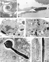
Figure 1
Figure 1
TEM of phagocytosis of C. albicans by DCs. FSDCs were incubated with C. albicans yeasts (A, C, and D) or hyphae (B, E, F, and G) at an FSDC/C. albicans ratio of 2:1, for 15 min (A and B), 1 h (E–G), 2 h (C), and 4 h (D) before processing for TEM. (A) Yeast engulfment through coiling phagocytosis and (B) hypha uptake through zipper-type phagocytosis, at 15 min after infection. (C) Fungal elements inside phagolysosomes at 2 h after exposure (arrows), and (D) in partially degraded forms at 4 h after exposure (arrow). (E) Hyphae escaping the phagosome at 2 h, and (F and G) lying free in the cytoplasm. Original magnifications: (A) ×17,000; (B) ×12,000; (C) ×7,000; (D) ×22,000; (E) ×12,000; (F) ×12,000; and (G) ×70,000. Bar, (A–C, E, F) 1 μm; (D) 0.5 μm; and (G) 0.1 μm.
Figure 3
Figure 3
Antifungal activities of DCs. (A) FSDCs were exposed to C. albicans yeasts or hyphae (FSDC/Candida ratios of 10:1, white bars; 5:1, hatched bars; and 2.5:1, black bars) for 2 h before determination of CFU inhibition, as described in Materials and Methods. (B) NO production by DCs unexposed (None), or upon exposure to IFN-γ (400 U/ml) and LPS (40 ng/ml) or Candida yeasts or hyphae. *P < 0.05, hypha-pulsed vs. yeast-pulsed DCs.
Figure 2
Figure 2
Effect of mannan on internalization of C. albicans by DCs. FSDCs were incubated with mannan at different concentrations for 10 min before the addition of C. albicans yeasts (white bars) or hyphae (black bars) at a ratio of 2:1 (FSDC/Candida). Internalized fungal cells were visualized by light microscopy after 30 min of incubation, as described in Materials and Methods. *P < 0.05, yeast or hypha internalization in the presence of mannan vs. yeast or hypha internalization in the absence of mannan.
Figure 4
Figure 4
Cytokine gene expression by DCs after exposure of C. albicans yeasts or hyphae. FSDCs were either unexposed (none) or exposed to fungal cells at a ratio of 2:1 for different time periods before analysis by RT-PCR. C, HPRT- or cytokine-specific control; N, no DNA added to the amplification mix during PCR.
Figure 5
Figure 5
Frequency of IL-4–producing cells in DCs exposed to C. albicans. FSDCs were exposed to yeasts or hyphae, at a ratio of 2:1, for 18 h before ELISPOT assay.
Figure 7
Figure 7
Intracellular cytokine expression in CD4+ T cells from BALB/c mice upon adoptive transfer of Candida-pulsed DCs. Splenic DCs, unpulsed (A) or ex vivo pulsed with Candida yeasts (B) or hyphae (C), were subcutaneously injected twice, once a week, 7 d before collection of splenocytes. For intracellular staining, purified CD4+ splenocytes were restimulated in vitro with APCs and inactivated Candida cells for 72 h. After washing, the cells were further stimulated with PMA and ionomycin in the presence of brefeldin A and then stained for intracellular IFN-γ and IL-4. Percentages reflect cytokine-positive cells.
Figure 6
Figure 6
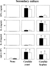
In vitro activation of CD4+ T cells by DCs upon exposure to C. albicans. FSDCs (white bars) or purified splenic DCs (black bars), either unexposed (none) or exposed to Candida yeasts or hyphae were cocultured with responder CD4+ T cells from naive B6D2F1 (for FSDCs) or BALB/c (for purified splenic DCs) mice (see Materials and Methods for details). After 5 d, cells were restimulated with fresh APCs (irradiated, T-depleted splenocytes) and inactivated Candida cells for 72 h (for cytokine determination) or 96 h (for lymphoproliferation). Cytokines were determined by specific ELISA (mean ± SE, ng/ml). Lymphoproliferation was measured by evaluating thymidine incorporation. The results are expressed as mean cpm ± SE of stimulated cells, after subtraction of cpm of unstimulated cells. *P < 0.05, Candida-pulsed vs. unpulsed FSDCs or DCs.
Figure 9
Figure 9
Effect of C. albicans–pulsed DCs from IL-12 KO or IL-4 KO mice on Th cell priming in vitro and in vivo. Purified DCs from spleens of mice with IL-12 p40 or IL-4 deficiency were ex vivo pulsed with Candida yeasts or hyphae, respectively, and cocultured in vitro with purified CD4+ T splenocytes from WT mice or adoptively transferred into WT recipients. Cocultures and cytokine determination were done as in the legend to Fig. 6. Infection and quantification of fungal growth in vivo were done as in the legend to Fig. 8. *P < 0.05, KO vs. WT mice.
Figure 8
Figure 8
Effect of adoptively transferred DCs on C. albicans growth and parameters of Th cell activation in mice with systemic candidiasis. Splenic DCs, unpulsed (white bars) or ex vivo pulsed with Candida yeast (black bars) or hyphae (hatched bars) were subcutaneously injected twice, once a week, 7 d before intravenous infection with C. albicans virulent strain. 7 d after infection, quantification of fungal growth was done in the kidneys, cytokines were determined by specific ELISA in supernatants of antigen-activated splenocytes, and lymphoproliferation of CD4+ T cells was assessed as in the legend to Fig. 6. Cross-hatched bars, mice vaccinated with the low-virulence C. albicans PCA-2, 2 wk before infection with virulent C. albicans. *P < 0.05, Candida-pulsed DCs or vaccinated mice vs. unpulsed DCs.

References

    1. Edwards J.E. Invasive Candida infectionsevolution of a fungal pathogen. N. Engl. J. Med. 1991;324:1060–1062. - PubMed
    1. Romani L., Puccetti P., Bistoni F. Biological role of helper T-cell subsets in candidiasis. Chem. Immunol. 1996;63:113–137. - PubMed
    1. Romani L. The T cell response to fungi. Curr. Opin. Immunol. 1997;9:484–490. - PubMed
    1. Romani L. Immunity to Candida albicansTh1, Th2 and beyond. Curr. Opin. Microbiol. 1999;2:363–367. - PubMed
    1. Lilic D., Cant A.J., Abinun M., Calvert J.E., Spickett G.P. Chronic mucocutaneous candidiasis. I. Altered antigen-stimulated IL-2, IL-4, IL-6 and interferon-gamma (IFN-γ) production. Clin. Exp. Immunol. 1996;5:205–212. - PMC - PubMed

Publication types

MeSH terms