

Human nerve growth factor protects common marmosets against autoimmune encephalomyelitis by switching the balance of T helper cell type 1 and 2 cytokines within the central nervous system
- PMID: 10811872
- PMCID: PMC2193155
- DOI: 10.1084/jem.191.10.1799
Human nerve growth factor protects common marmosets against autoimmune encephalomyelitis by switching the balance of T helper cell type 1 and 2 cytokines within the central nervous system
Abstract
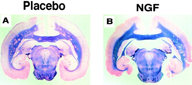
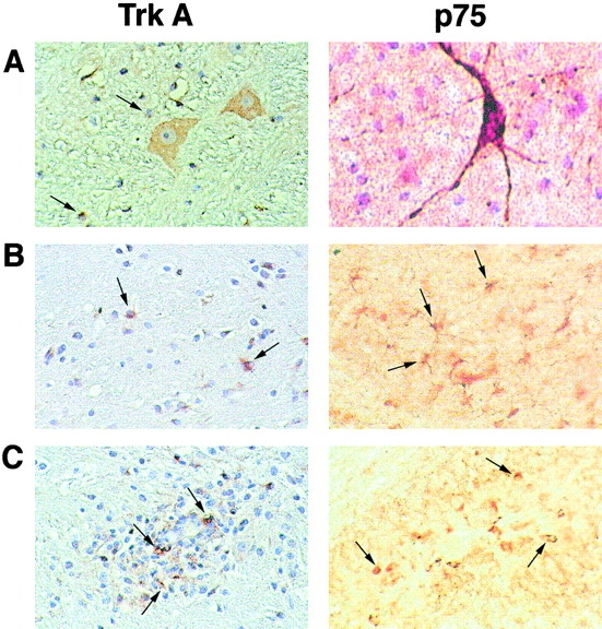
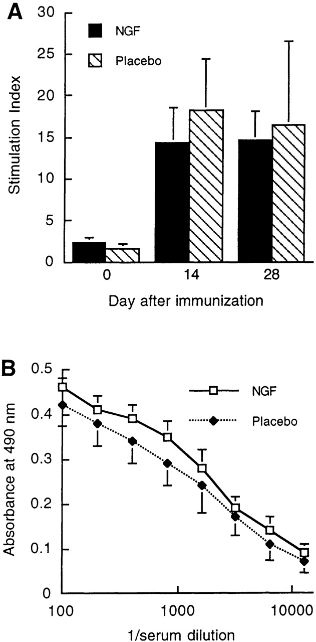
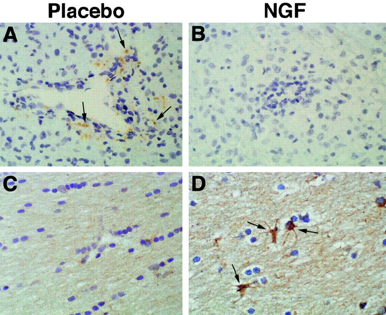
Multiple sclerosis is a demyelinating disorder of the central nervous system (CNS), in which an immune attack directed against myelin constituents causes myelin destruction and death of oligodendrocytes, the myelin-producing cells. Here, the efficacy of nerve growth factor (NGF), a growth factor for neurons and oligodendrocytes, in promoting myelin repair was evaluated using the demyelinating model of experimental allergic encephalomyelitis (EAE) in the common marmoset. Surprisingly, we found that NGF delayed the onset of clinical EAE and, pathologically, prevented the full development of EAE lesions. We demonstrate by immunocytochemistry that NGF exerts its antiinflammatory effect by downregulating the production of interferon gamma by T cells infiltrating the CNS, and upregulating the production of interleukin 10 by glial cells in both inflammatory lesions of EAE and normal-appearing CNS white matter. Thus, NGF, currently under investigation in human clinical trials as a neuronal trophic factor, may be an attractive candidate for therapy of autoimmune demyelinating disorders.
Figures
Comment in
-
Surprising pleiotropy of nerve growth factor in the treatment of experimental autoimmune encephalomyelitis.J Exp Med. 2000 May 15;191(10):1625-30. doi: 10.1084/jem.191.10.1625. J Exp Med. 2000. PMID: 10811856 Free PMC article. Review. No abstract available.
References
-
- Raine C. Demyelinating diseases. In: Davis R., Robertson D., editors. Textbook of Neuropathology. 3rd ed. Williams & Wilkins; Baltimore: 1997. pp. 627–714.
-
- Hohlfeld R. Biotechnological agents for the immunotherapy of multiple sclerosis. Principles, problems and perspectives. Brain. 1997;120:865–916. - PubMed
-
- Althaus H., Kloppner S., Schmidt-Schultz T., Schwartz P. Nerve growth factor induces proliferation and enhances fiber regeneration in oligodendrocytes isolated from adult pig brain. Neurosci. Lett. 1992;135:219–223. - PubMed
-
- Urschel B., Hulsebosch C. Schwann cell-neuronal interactions in the rat involve nerve growth factor. J. Comp. Neurol. 1990;296:114–122. - PubMed
Publication types
MeSH terms
Substances
Grants and funding
LinkOut - more resources
Full Text Sources
Other Literature Sources
