Review
. 2000 Mar;74(2-3):291-7.
doi: 10.1016/s0031-6865(99)00056-4.

ICAM-1 receptors and cold viruses

Affiliations
Review

ICAM-1 receptors and cold viruses

J Bella et al. Pharm Acta Helv. 2000 Mar.

Abstract

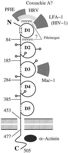
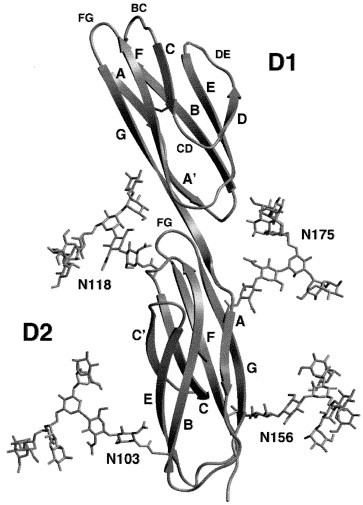
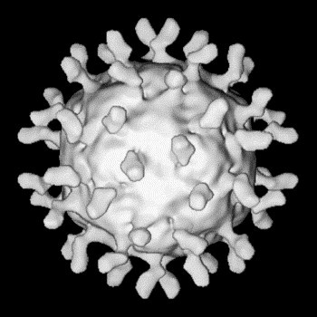
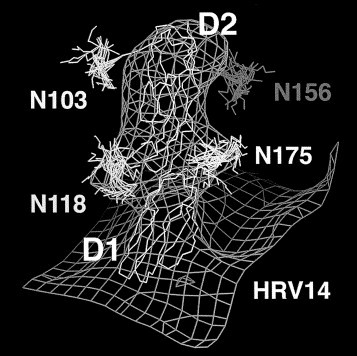
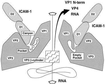
Human rhinoviruses (HRVs), the single most important etiologic agent of common colds, are small viruses composed of an icosahedral protein shell that encapsidates a single, positive RNA strand. Multiplication of HRVs occurs in the cytoplasm of the host cell. To produce infection, HRVs must first attach to specific cellular receptors embedded in the plasma membrane. Ninety percent of HRVs immunogenic variants use as receptor intercellular adhesion molecule-1 (ICAM-1), a cell surface glycoprotein that promotes intercellular signaling in processes derived from inflammation response. As HRV receptor, ICAM-1 positions the virus to within striking distance of the membrane, and then triggers a conformational change in the virus that ultimately results in delivery of the viral RNA genome into the cytoplasm, across a lipid bilayer. The interaction between ICAM-1 and HRVs has been analyzed by the combination of crystal structures of HRVs and ICAM-1 fragments with electron microscopy reconstructions of the complexes. The resulting molecular models are useful to address questions about receptor recognition, binding specificity, and mechanisms by which ICAM-1 induces virus uncoating.


Figures

Fig. 1
Fig. 1
Domain structure of ICAM-1. Each Ig domain is represented schematically by a circle closed by one or two disulfide bonds. Amino acid numbers indicate the beginning and end of each domain. Approximate locations of relevant binding sites are shown. Lollipop-shaped structures indicate N-linked glycosylation sites.
Fig. 2
Fig. 2
Ribbon diagram of the first two domains of ICAM-1. Strands are labeled A, B, C, etc., following the convention for Ig domain nomenclature (Harpaz and Chothia, 1994). Loops are named by the strands they connect. Only functionally relevant loops mentioned in the text are labeled. Glycan models show the position of the four glycosylation sites in domain D2, linked to four asparagine residues. Only the first one or two sugars in each glycan can be identified in ICAM-1 D1D2 crystal structures (Bella et al., 1998; Casasnovas et al., 1998).
Fig. 3
Fig. 3
Cryo-EM reconstruction of a complex between HRV16 and a low-glycosylation form of ICAM-1 D1D2 (Bella et al., 1998). There are 60 copies of the receptor fragment, visible as radial projections on the viral surface.
Fig. 4
Fig. 4
Fitting of an atomic model for ICAM-1 D1D2 into the cryo-EM reconstruction of its complex with HRV14. Protein and carbohydrate are represented as Cα and C1–C4 tracings, respectively.
Fig. 5
Fig. 5
Scheme of a two-step binding mechanism between ICAM-1 and major group HRVs. The first step, on the left, is observed in the cryo-EM reconstructions of HRV-ICAM-1 fragments. On the right, the second (hypothesized) step involves a conformational change in the virus surface. The five-fold channel may open as both walls and floor of the canyon bind to domain D1 of ICAM-1. The pocket region is postulated as the hinge area, and needs to be empty in order to provide conformational flexibility.

Similar articles

Cited by

References

    1. Bastiani L, Laal S, Kim M, Zolla-Pazner S. Host cell-dependent alterations in envelope components of human immunodeficiency virus type 1 virions. J. Virol. 1997;71:3444–3450. - PMC - PubMed
    1. Bella J, Kolatkar P.R, Marlor C.W, Greve J.M, Rossmann M.G. The structure of the two amino-terminal domains of human ICAM-1 suggests how it functions as a rhinovirus receptor and as an LFA-1 integrin ligand. Proc. Natl. Acad. Sci. U. S. A. 1998;95:4140–4145. - PMC - PubMed
    1. Berendt A.R, McDowall A, Craig A.G, Bates P.A, Sternberg M.J.E, Marsh K, Newbold C.I, Hogg N. The binding site on ICAM-1 for Plasmodium falciparum-infected erythrocytes overlaps, but is distinct from, the LFA-1-binding site. Cell. 1992;68:71–81. - PubMed
    1. Carpén O, Pallai P, Staunton D.E, Springer T.A. Association of intercellular adhesion molecule-1 (ICAM-1) with actin-containing cytoskeleton and α-actinin. J. Cell Biol. 1992;118:1223–1234. - PMC - PubMed
    1. Casasnovas J.M, Springer T.A, Liu J.-H, Harrison S.C, Wang J.-H. Crystal structure of ICAM-2 reveals a distinctive integrin recognition surface. Nature. 1997;387:312–315. - PubMed

Substances

LinkOut - more resources