Comparative Study
. 2000 May;21(5):852-61.

Early MR features of hypoxic-ischemic brain injury in neonates with periventricular densities on sonograms

Affiliations
Comparative Study

Early MR features of hypoxic-ischemic brain injury in neonates with periventricular densities on sonograms

L T Sie et al. AJNR Am J Neuroradiol. 2000 May.

Abstract

Background and purpose: In the early 1980s, diagnosing periventricular leukomalacia (PVL) in neonates by using cranial sonography was possible for the first time. Our purpose was to investigate the possibility of diagnosing PVL in the acute stage by using MR imaging. We evaluated early MR features of hypoxic-ischemic brain injury in neonates with periventricular densities (flares) on cranial sonograms to determine the added value of MR imaging over sonography alone for early diagnosis of brain damage.

Methods: In a prospective study, infants who showed flares and/or cysts on sonograms underwent MR imaging during the (sub)acute stage.

Results: Fifty infants were classified according to the highest sonographic grade up to the day of MR imaging: 23 infants had sonographic grade 1 (flares < 1 week), 15 had sonographic grade 2 (flares > or = 1 week), four had sonographic grade 3 (small localized cysts), and eight had sonographic grade 4 (extensive periventricular cysts); none had sonographic grade 5 (multicystic leukomalacia) on the day of MR imaging. Overall, the additional information provided by MR imaging (over sonography alone) consisted of the depiction of hemorrhagic lesions in 64% of the infants. Extent and severity of the hemorrhages varied from isolated punctate lesions to extensive hemorrhages throughout the white matter; the latter were followed by cystic degeneration at autopsy in two infants. In nine of the 12 infants with cystic PVL, MR images showed more numerous or more extensive cysts. In addition, in two infants, MR images showed cysts not present on sonograms. In 32% of the infants, MR imaging provided no additional information; in these children, all but one had flares on sonograms whereas MR images showed no abnormalities or a zone of mild periventricular signal change.

Conclusion: MR imaging can depict the precise site and extent of hypoxic-ischemic brain injury at an earlier stage and allows a wider differentiation of lesions as compared with sonography alone. Hemorrhagic PVL is considered to be rare, but was present in 64% of our study population.


Figures

None
FIG 1. Neonatal MR score versus initial sonographic (US) score (n = 50). Solid circles represent infants with the same initial and end sonographic score; open circles, infants with a difference between initial and end sonographic scores
None
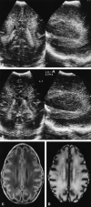
FIG 2. Infant born at 28 4/7 weeks. A–D, Transient homogeneous periventricular densities are seen in the frontoparietal region on coronal and parasagittal sonograms at a PCA of 29 2/7 weeks (A). Coronal and parasagittal sonograms at a PCA of 34 4/7 weeks (B) show normalized echogenicity of the white matter with a homogeneous aspect. Axial T1-weighted (C) and T2-weighted (D) MR images on the same day show a frontal and parietal periventricular zone of mildly changed signal intensity.
None
FIG 3. Infant born at 34 2/7 weeks. A–C, Parasagittal sonogram at a PCA of 35 5/7 weeks (A) shows periventricular densities with an inhomogeneous aspect in the frontoparietal region. T1-weighted MR images on the same day in the parasagittal (B) and axial (C) planes show multiple, small, punctate hemorrhages, occurring in the periventricular white matter on both sides.
None
FIG 4. Infant born at 37 3/7 weeks. A–D, Coronal and parasagittal sonograms (A) at a PCA of 38 2/7 weeks show a diffuse increased echogenicity throughout the white matter and basal ganglia with an inhomogeneous, partly precystic aspect in the frontal region. Proton density–weighted (B), T2-weighted (C), and inversion recovery (D) MR images in the axial plane show large areas of high signal on the proton density–weighted image (B) and combined high and low signal changes on the T2-weighted and inversion recovery images throughout the white matter (C, D) and basal ganglia (D). In comparison with sonograms, MR images show more extensive, small, partly cystic and partly precystic periventricular lesions in the frontal and parietal region, with signal intensities identical or close to CSF (B–D). The hemorrhagic ischemic areas correspond to the large cystic lesions detected at autopsy performed 4 days later.
None
FIG 5. Infant born at 37 6/7 weeks. A and B, Coronal sonogram (A) at a PCA of 39 weeks shows a strikingly asymmetrical increased echogenicity of the periventricular white matter, predominantly in the left parietal region, with an inhomogeneous aspect. In addition, intraventricular and germinal-layer hemorrhages were present. T1-weighted MR image on the same day in the axial plane (B) shows extensive signal intensity changes and hemorrhagic lesions throughout the left hemisphere and a small periventricular hemorrhage on the other side.
None
FIG 6. Infant born at 32 weeks. A and B, Parasagittal sonogram (A) at a PCA of 33 4/7 weeks shows a (stable) posthemorrhagic ventricular dilatation and a moderately increased echogenicity of the white matter in the parietooccipital region with an inhomogeneous aspect and few small cystic lesions. Axial T1-weighted MR image on the same day (B) shows extensive white matter signal intensity changes with multiple hemorrhagic lesions and more numerous cystic lesions.
None
FIG 7. Infant born at 34 5/7 weeks. A and B, Parasagittal (A) and coronal (B) sonograms at a PCA of 36 5/7 weeks show a moderately increased echogenicity of the periventricular white matter with a homogeneous aspect and one large cyst in the parietal region. Axial T1-weighted MR image on the same day (C) shows more numerous, bilateral, large periventricular cysts on MR images, with focal extension into the subcortical white matter in the entire parietooccipital region.
None
FIG 8. Neonatal MR score versus sonographic (US) end score (n = 48). Solid circles represent infants with the same initial and end sonographic score; open circles, infants with a difference between initial and end sonographic scores; cross, infants who did not survive the neonatal period

References

    1. Hill A, Melson GL, Clark HB, Volpe JJ. Hemorrhagic periventricular leukomalacia: diagnosis by real time ultrasound and correlation with autopsy findings. Pediatrics 1982;69:282-284 - PubMed
    1. De Vries LS, Eken P, Dubowitz LMS. The spectrum of leukomalacia using cranial ultrasound. Behav Brain Res 1992;49:1-6 - PubMed
    1. Hope PL, Gould SJ, Howard S, Hamilton PA, Costello AM de L, Reynolds EOR. Precision of ultrasound diagnosis of pathologically verified lesions in the brains of very preterm infants. Dev Med Child Neurol 1988;30:457-471 - PubMed
    1. Carson SC, Hertzberg BS, Bowie JD, Burger PC. Value of sonography in the diagnosis of intracranial hemorrhage and periventricular leukomalacia: a postmortem study of 35 cases. AJNR Am J Neuroradiol 1990;11:677-683 - PMC - PubMed
    1. Eken P, Jansen GH, Groenendaal F, Rademaker KJ, De Vries LS. Intracranial lesions in the fullterm infant with hypoxic ischaemic encephalopathy: ultrasound and autopsy correlation. Neuropediatrics 1994;25:301-307 - PubMed

Publication types