

Multiple sclerosis: comparison of trace apparent diffusion coefficients with MR enhancement pattern of lesions
- PMID: 10815662
- PMCID: PMC7976762
Multiple sclerosis: comparison of trace apparent diffusion coefficients with MR enhancement pattern of lesions
Abstract
Background and purpose: Diffusion-weighted MR imaging and the trace apparent diffusion coefficient (ADC) provide important structural information about tissues. The purpose of this study was to investigate the relationship between trace ADC values and the enhancement pattern of multiple sclerosis (MS) lesions.
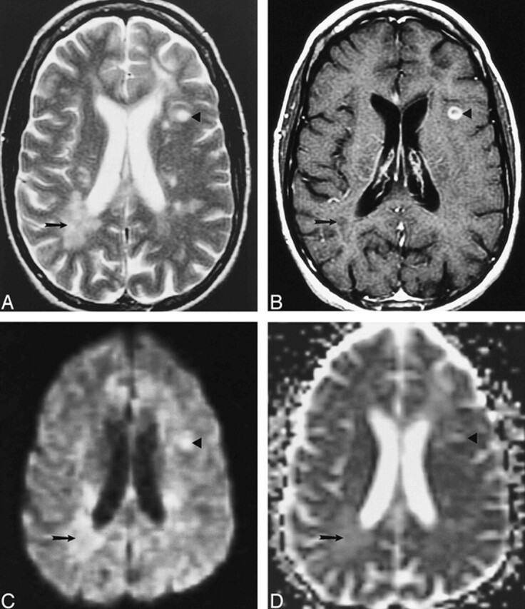
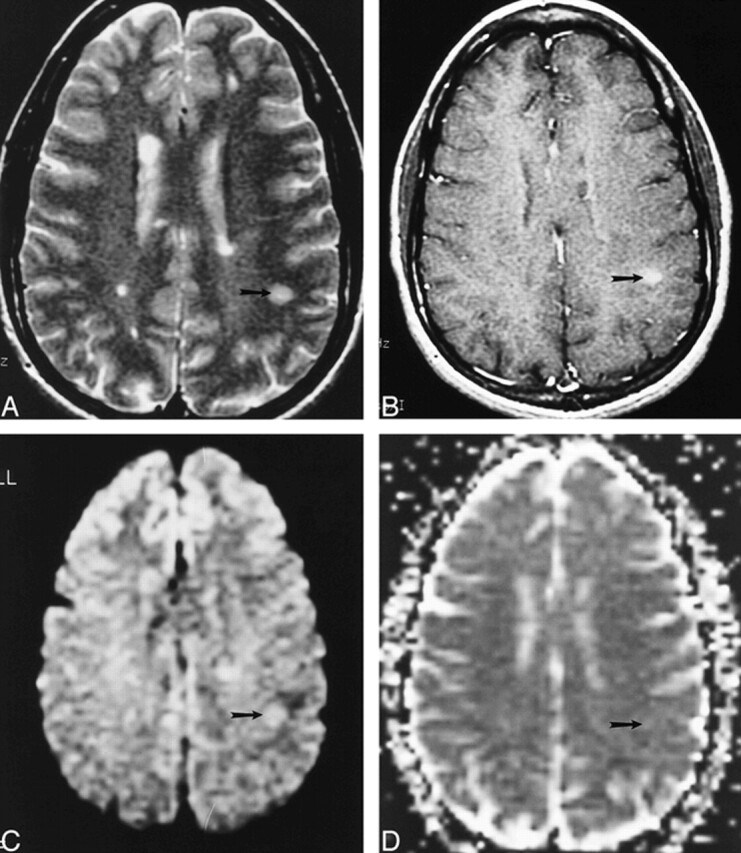
Methods: Ninety-six lesions, identified in 24 patients with MS, were characterized by their enhancement pattern on contrast-enhanced T1-weighted MR images. There were 57 nonenhancing lesions (NELs), 28 homogeneously enhancing lesions (HELs), and 11 ring-enhancing lesions (RELs). The trace ADC means for each type of lesion and for normal-appearing white matter (NAWM) were calculated and compared using Student's t-test.
Results: The mean trace ADC values for HELs (mean, 7.7 x 1(-10) m2s(-1); SD, 1.4 x 10(-10) m2s(-1)) were less than those for RELs (mean, 1.2 x 10(-9) m2s(-1); SD, 3.5 x 10(-10)m2s(-1)) and NELs (mean, 1.3 x 10(-9) m2(s-1); SD, 2.6 x 10(-10) m2(s-1)). There was a significant difference between the mean trace ADC values of HELs and RELs as well as between those for HELs and NELs. There was also a significant difference in the mean trace ADC values between all lesion types and NAWM (mean, 6.9 x 10(-10) m2s(-1); SD, 5.0 x 10(-11) m2s(-1)).
Conclusion: We found a predictable relationship between mean trace ADC and the pattern of enhancement in MS lesions, corresponding to reported histopathologic differences in myelination between lesion types and magnetization transfer ratios.
Figures
References
-
- Katz D, Taubenberger J, Cannella B, McFarlin D, Raine C, McFarland H. Correlation between magnetic resonance imaging findings and lesion development in chronic, active multiple sclerosis. Ann Neurol 1993;34:661-669 - PubMed
-
- Nesbit G, Forbes G, Scheithauer B, Okazaki H, Rodriguez M. Multiple sclerosis: histopathological and MR and/or CT correlation in 37 cases at biopsy and three cases at autopsy. Radiology 1991;180:467-474 - PubMed
-
- vanWalderveen M, Kamphorst W, Scheltens P, et al. Histopathologic correlate of hypointense lesions on T1-weighted spin-echo MRI in multiple sclerosis. Neurology 1998;50:1282-1288 - PubMed
-
- Stejskal E, Tanner J. Spin diffusion measurements: spin echoes in the presence of a time-dependent field gradient. J Chem Phys 1965;42:288-292
Publication types
MeSH terms
Substances
Grants and funding
LinkOut - more resources
Full Text Sources
Medical