. 2000 Jun;68(6):3108-15.
doi: 10.1128/IAI.68.6.3108-3115.2000.

Enterohemorrhagic Escherichia coli induces apoptosis which augments bacterial binding and phosphatidylethanolamine exposure on the plasma membrane outer leaflet

Affiliations

Enterohemorrhagic Escherichia coli induces apoptosis which augments bacterial binding and phosphatidylethanolamine exposure on the plasma membrane outer leaflet

D Barnett Foster et al. Infect Immun. 2000 Jun.

Abstract

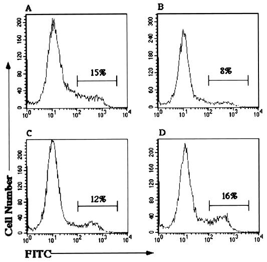
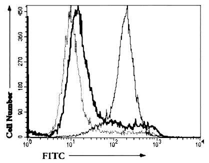
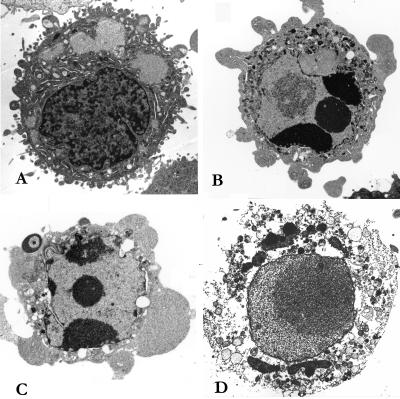
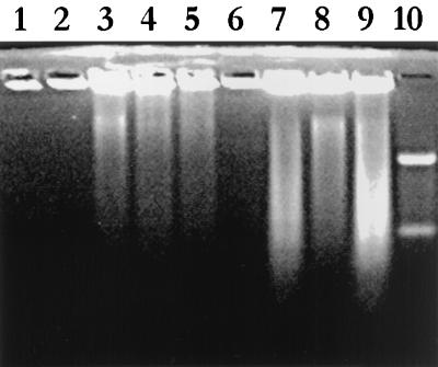
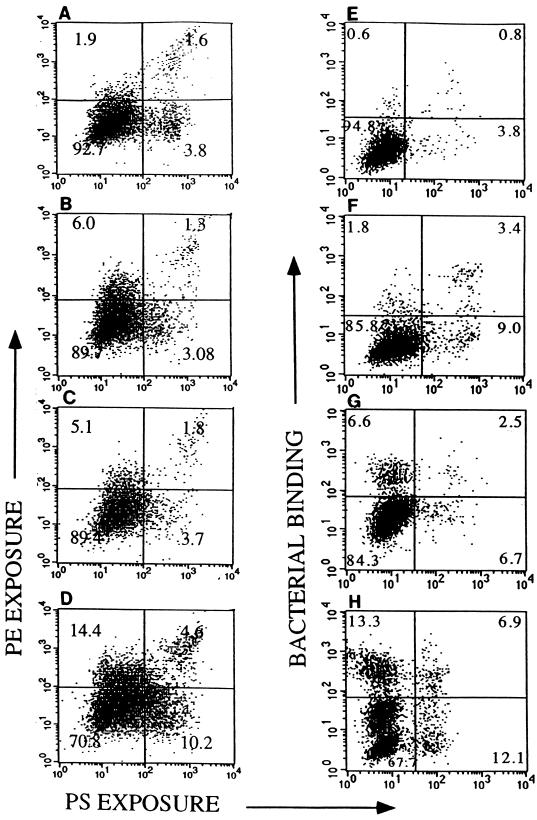
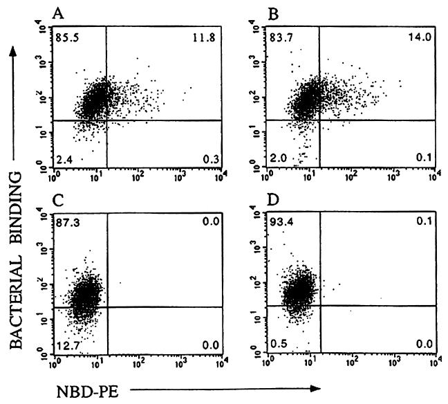
Enterohemorrhagic Escherichia coli (EHEC) is a gastrointestinal pathogen that causes watery diarrhea and hemorrhagic colitis and can lead to serious and even fatal complications such as hemolytic uremic syndrome. We investigated the ability of EHEC to kill host cells using three human epithelial cell lines. Analysis of phosphatidylserine expression, internucleosomal cleavage of host cell DNA and morphological changes detected by electron microscopy changes revealed evidence of apoptotic cell death. The rates and extents of cell death were similar for both verotoxin-producing and nonproducing strains of EHEC as well as for a related gastrointestinal pathogen, enteropathogenic E. coli (EPEC). The induction of apoptosis by bacterial attachment was independent of verotoxin production and greater than that produced by a similar treatment with verotoxin alone. Expression of phosphatidylethanolamine, previously reported to bind EHEC and EPEC, was also increased on apoptotic cells but with little correlation to phosphatidylserine expression. Phosphatidylethanolamine levels but not phosphatidylserine levels on dying cells correlated with EHEC binding. Cells treated with phosphatidylethanolamine-containing liposomes also showed increased EHEC binding. These results suggest that bacterial induction of apoptosis offers an advantage for bacterial attachment by augmenting outer leaflet levels of the phosphatidylethanolamine receptor.


Figures

FIG. 1
FIG. 1
Flow cytometric analysis of outer leaflet levels of PS in HEp-2 cells. The x axis represents the staining intensity on the cells; the y axis represents the relative cell number. The percentage of cells exhibiting increased fluorescence intensity is indicated. Cells were preincubated with the following: (A) VT1 (12.5 ng/ml overnight) or 108 bacteria for 5 h; (B) HB101; (C) EPEC (E2348/69); (D) EHEC (85-170). Membrane PS was detected using annexin V-FITC. Incubation with EHEC 84-289 and CL56 produced histograms similar to panel D; PS levels for HEp-2 cells alone produced results similar to panel B.
FIG. 2
FIG. 2
Effect of bacterial incubation time on PS expression. HEp-2 cells were incubated with EHEC (85-170) for 2 h (dotted), 3 h (black), or overnight (grey). PS expression was determined by flow cytometric analysis of annexin V-FITC binding as for Fig. 1. Similar results (not shown) were achieved with EHEC strains 84-289 and CL56.
FIG. 3
FIG. 3
Electron micrographs of HEp-2 cells. (A) Viable cell (untreated); (B) apoptotic cell (treated with VT1 [12.5 ng/ml] overnight); (C) apoptotic cell (treated with CL56 for 5 h); (D) necrotic cell (treated with VT1 [12.5 ng/ml] overnight).
FIG. 4
FIG. 4
Analysis of internucleosomal DNA fragmentation by agarose gel electrophoresis of DNA extracts from HEp-2 cells incubated with medium (lane 1), 100 ng of VT1 (24 h) (lane 2), 100 ng of VT1 (48 h) (lane 3), 200 ng of VT1 (24 h) (lane 4), 200 ng of VT1 (48 h) (lane 5), HB101 (5 h) (lane 6), 85-170 (5 h) (lane 7), 84-289 (5 h) (lane 8), or E2348/69 (5 h) (lane 9). Lane 10, 100-kb standard.
FIG. 5
FIG. 5
Flow cytometric analysis of PE and PS exposure and EHEC binding to HEp-2. Cells were preincubated for various times (0 to 20 h) with 12.5 ng of VT1 per ml in serum-free media. (A to D) Cells treated both with annexin V-FITC (to detect PS) and anti-PE (visualized with phycoerythrin-GAR conjugate) to compare PS (abscissa) and PE (ordinate) exposure with VT1 incubation time. (A) No VT1; (B) VT1, 1 h; (C) VT1, 3 h; (D) VT1, 20 h. (E to H) Cells pretreated with VT1 or medium, infected with 108 CL56, and stained with annexin V-FITC and a bacterium-specific antiserum to compare PS exposure (abscissa) with bacterial binding (ordinate). (E) Control (no VT1, no CL56); (F) VT1, 20 h (no CL56); (G) CL56 (no VT); (H) VT1 (20 h), CL56. Quadrant numbers refer to percentage of cells simultaneously stained with both fluorophores with intensities as indicated on the axes.
FIG. 6
FIG. 6
Bacterial binding versus NBD-PE uptake. Flow cytometric dot plots show the fluorescence intensity of NBD-PE staining on the abscissa and bacterial binding (FITC staining) on the ordinate. Bacterial binding was determined for CL56 with cells treated with NBD-PE liposomes (A), cells pretreated with NEM and then NBD-PE liposomes (B), untreated cells (C), and cells pretreated with NEM (D). Additional controls (not shown) included cells incubated with primary and/or secondary antibody and cells incubated only with NBD-PE liposomes.

References

    1. Acheson D W K, Moore R, De Breucker S, Lincicome L, Jacewicz M, Skutelsky E, Keusch G T. Translocation of Shiga toxin across polarized intestinal cells in tissue culture. Infect Immun. 1996;64:3294–3300. - PMC - PubMed
    1. Barnett Foster D E, Philpott D, Abul-Milh M, Huesca M, Sherman P M, Lingwood C A. Phosphatidylethanolamine recognition promotes enteropathogenic and enterohemorrhagic E. coli host cell attachment. Microb Pathog. 1999;27:289–301. - PubMed
    1. Bauer M E, Welsh R A. Characterization of an RTX toxin from enterohemorrhagic Escherichia coli O157:H7. Infect Immun. 1996;64:167–175. - PMC - PubMed
    1. Björk S, Breimer M E, Hansson G C, Karlsson K-A, Leffler H. Structures of blood group glycosphingolipids in human small intestine. J Biol Chem. 1987;262:6758–6765. - PubMed
    1. Boyd B, Lingwood C A. Verotoxin receptor glycolipid in human renal tissue. Nephron. 1989;51:207–210. - PubMed

Publication types

Substances

LinkOut - more resources