. 2000 Jun;231(6):838-48.
doi: 10.1097/00000658-200006000-00008.

Gut peptide receptor expression in human pancreatic cancers

Affiliations

Gut peptide receptor expression in human pancreatic cancers

R A Ehlers et al. Ann Surg. 2000 Jun.

Abstract

Objective: To determine the prevalence of gastrointestinal (GI) peptide receptor expression in pancreatic cancers, and to further assess signaling mechanisms regulating neurotensin (NT)-mediated pancreatic cancer growth.

Summary background data: Pancreatic cancer remains one of the leading causes of GI cancer death; novel strategies for the early detection and treatment of these cancers is required. Previously, the authors have shown that NT, an important GI hormone, stimulates the proliferation of an NT receptor (NTR)-positive pancreatic cancer.

Methods: A total of 26 human pancreatic adenocarcinomas, obtained after resection, and 5 pancreatic cancer xenografts were analyzed for expression of NTR, vasoactive intestinal peptide receptor (VIPR), substance P receptor (SPR), and gastrin-releasing peptide receptor (GRPR). In addition, NTR expression, [Ca2+]i mobilization, and growth in response to NT was determined in L3.6, a metastatic pancreatic cancer cell line.

Results: Neurotensin receptor was expressed in 88% of the surgical specimens examined and all five of the pancreatic cancer xenografts. In contrast, VIPR, SPR, and GRPR expression was detected in 31%, 27%, and 8% of pancreatic cancers examined, respectively. Expression of NTR, functionally coupled to the Ca2+ signaling pathway, was identified in L3.6 cells; treatment with NT (10 micromol/L) stimulated proliferation of these cells.

Conclusions: The authors demonstrated NTR expression in most of the pancreatic adenocarcinomas examined. In contrast, VIPR, SPR, and GRPR expression was detected in fewer of the pancreatic cancers. The expression of NTR and other peptide receptors suggests the potential role of endocrine manipulation in the treatment of these cancers. Further, the presence of GI receptors may provide for targeted chemotherapy or radiation therapy or in vivo scintigraphy for early detection.


Figures

None
Figure 1. Gut peptide receptor expression in human pancreatic cancers. RNA was extracted from pancreatic cancer specimens resected from patients (n = 12), and reverse transcriptase—polymerase chain reaction (RT-PCR) was performed to determine expression of neurotensin receptor (NTR), vasoactive intestinal peptide receptor (VIPR), substance P receptor (SPR), and gastrin-releasing peptide receptor (GRPR). To confirm integrity of the RT reaction, primers for the constitutively active gene GAPDH were used for amplification (lower panel).
None
Figure 2. Gut peptide receptor expression in human pancreatic cancer xenografts. RNA was extracted from five pancreatic cancer xenografts established in athymic nude mice from resected pancreatic cancers. Reverse transcriptase—polymerase chain reaction (RT-PCR) was performed to detect expression of neurotensin receptor (NTR), vasoactive intestinal peptide receptor (VIPR), substance P receptor (SPR), and gastrin-releasing peptide receptor (GRPR). In addition, the integrity of the RT reaction was confirmed by amplification using primers to the constitutively expressed GAPDH gene.
None
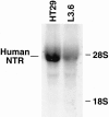
Figure 3. Neurotensin receptor (NTR) is expressed in L3.6 pancreatic cancer cells. Northern blot analysis of total RNA (50 μg) from L3.6 pancreatic cancer cells. RNA extracted from the HT29 cell line was used as positive control for NTR expression. Blots were probed with a 32P-labeled human NTR cDNA probe.
None
Figure 4. Intracellular calcium ([Ca2+]i) mobilization in L3.6 pancreatic cancer cells. (A) Pseudocolor images representing the relative [Ca2+]i concentration as demonstrated by the color bar (left). Times listed are after treatment with neurotensin (NT; 50 nM). (B) Graph depicting [Ca2+]i concentration over a time course after NT treatment (50 nM). Each line represents a single cell measurement. To ensure the ability of cells to reproduce a calcium response, NT treatment was followed with acetylcholine (Ach) treatment (100 mmol/L). (C) Graph depicting [Ca2+]i concentration in L3.6 cells pretreated with SR48692 (1 mmol/L) before NT treatment (50 nM). To verify the specificity of inhibition of the NT receptor-mediated calcium response, cells were treated with Ach (100 mmol/L).
None
Figure 5. Dose-response for neurotensin receptor (NTR) stimulation and inhibition in L3.6 pancreatic cancer cells. (A) Graph depicting maximal increase in [Ca2+]i in spectrum response to increasing doses of neurotensin (NT). Each data point represents the mean of multiple cell measurements ± standard error. (B) Graph depicting maximal increase in [Ca2+]i in cells pretreated with increasing doses of SR48692 followed by NT (50 nM). Data are expressed as percentage of increase compared with control cells treated with NT (50 nM) alone. Each data point represents the mean of multiple cell measurements ± standard error.
None
Figure 6. Neurotensin (NT) treatment increases growth of L3.6 pancreatic cancer cells. Graph depicting MTT absorbance at 570 nm in L3.6 cells treated with increasing doses of NT. MTT assays were performed on days 4 and 6. Data are expressed as mean ± standard error. (*P < .05 vs. control [CON]).

References

    1. Landis SH, Murray T, Bolden S, Wingo PA. Cancer statistics, 1999. CA Cancer J Clin 1999; 49:8–31. - PubMed
    1. Scheithauer W, Kornek GV, Raderer M, et al. Phase II trial of gemcitabine, epirubicin and granulocyte colony-stimulating factor in patients with advanced pancreatic adenocarcinoma. Br J Cancer 1999; 80:1797–1802. - PMC - PubMed
    1. Royce ME, Pazdur R. Novel chemotherapeutic agents for gastrointestinal cancers. Curr Opin Oncol 1999; 11:299–304. - PubMed
    1. Green S, Furr B. Prospects for the treatment of endocrine-responsive tumours. Endocr Relat Cancer 1999; 6:349–371. - PubMed
    1. Townsend CM Jr, Singh P, Thompson JC. Hormonal effects on gastrointestinal cancer growth. In: Etievant C, Cros J, Rustum YM, eds. New Concepts in Cancer: Metastasis, Oncogenes and Growth Factors. London: MacMillan Press; 1990: 208–217.

Publication types

MeSH terms

Substances