. 2000 Jun;44(6):1518-23.
doi: 10.1128/AAC.44.6.1518-1523.2000.

Morphological changes and lysis induced by beta-lactams associated with the characteristic profiles of affinities of penicillin-binding proteins in actinobacillus pleuropneumoniae

Affiliations

Morphological changes and lysis induced by beta-lactams associated with the characteristic profiles of affinities of penicillin-binding proteins in actinobacillus pleuropneumoniae

T Inui et al. Antimicrob Agents Chemother. 2000 Jun.

Abstract

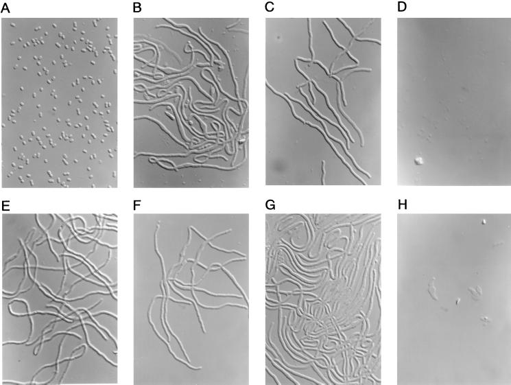
Actinobacillus pleuropneumoniae, which was formerly classified in the genus Haemophilus, is a pathogen causing swine pleuropneumonia. We found that aspoxicillin showed strong activity and that meropenem had better lytic activity against this pathogen. In the present study, we for the first time identified penicillin-binding proteins (PBPs) of A. pleuropneumoniae in order to elucidate the relationship between the antibacterial and lytic activities of beta-lactam antibiotics and affinities of the PBPs. The competitive assay using (3)H-labeled benzylpenicillin revealed seven PBPs in A. pleuropneumoniae; they were determined to be PBPs 1a, 1b, 2, 3, 4, 5, and 6, and the molecular masses of these PBPs were estimated to be 92, 80, 76, 72, 50, 44, and 30 kDa, respectively, by comparison with those of Haemophilus influenzae. Our detailed analysis of the affinities of the PBPs of A. pleuropneumoniae and of the bacterial lysis kinetics for several beta-lactam antibiotics revealed that the strong antibacterial activity of aspoxicillin against this strain could be related to the higher affinity of PBP 3 and that preferential inactivation of PBP 1b could cause rapid lysis.


Figures

FIG. 1
FIG. 1
Comparative profiles of PBPs of A. pleuropneumoniae NB001 and H. influenzae IID983. PBPs in the cell membrane preparations labeled with 3H-PCG were fractionated by 12.5% polyacrylamide gel electrophoresis and visualized with the BAS-2000 image-analyzing system. Lanes A and B show PBPs untreated and PBPs treated with PCG (100 μg/ml), respectively. The molecular mass (in kilodaltons) of each PBP, indicated in parentheses, of A. pleuropneumoniae NB001 was estimated from that of H. influenzae (12).
FIG. 2
FIG. 2
Time-kill (A) and lytic (B) studies on aspoxicillin against A. pleuropneumoniae NB001. Viable cell counts were determined by the colony-counting method, and the turbidity of the culture was represented as an OD620 value. Aspoxicillin was added to the culture at time zero at concentrations of its MIC (circles), 4× its MIC (triangles), and 16× its MIC (squares). The viable cell counts and the OD of a control culture at times of −1, 0, 1, 2, 4, and 8 h are represented by dotted lines without symbols.
FIG. 3
FIG. 3
Bacteriolytic kinetics of meropenem (A), cefsulodin (B), cefdinir (C), and piperacillin (D) against A. pleuropneumoniae NB001. Each drug was added to the culture at time zero in the exponential phase at concentrations of its MIC (circles), at 4× its MIC (squares), and at 16× the MIC (squares). The OD of a control culture at times of −1, 0, 1, 2, 4, and 8 h are represented by dotted lines without symbols.
FIG. 4
FIG. 4
Photomicrographs of A. pleuropneumoniae NB001 treated with each antibiotic at 4× the MIC for 2 h and are depicted as follows: none (A), aspoxicillin (B), piperacillin (C), mecillinam (D), aztreonam (E), cefdinir (F), cefsulodin (G), and meropenem (H). Magnification, ×1,000.
FIG. 5
FIG. 5
Time-kill (A) and lytic (B) studies with aspoxicillin and piperacillin against H. influenzae IID983. Viable cell counts were determined by the colony-counting method, and the turbidity of culture was represented as the OD620. Closed and opened symbols represent aspoxicillin and piperacillin, respectively. Each antibiotic was added to the culture at time zero at concentrations of its MIC (circles) and at 4× its MIC (triangles). The viable cell counts and the OD values of a control culture at times of −1, 0, 1, 2, 4, and 8 h are represented by dotted lines without symbols.
FIG. 6
FIG. 6
Photomicrographs of H. influenzae IID983 treated with aspoxicillin and piperacillin at 4× the MIC for 2 h are depicted as follows: none (A), aspoxicillin (B), and piperacillin (C). Magnification, ×1,000.

References

    1. Asawa T, Kobayashi H, Mitani K, Ito N, Morozumi T. Serotypes and antimicrobial susceptibility of Actinobacillus pleuropneumoniae isolated from piglets with pleuropneumonia. J Vet Med Sci. 1995;57:757–759. - PubMed
    1. Clairoux N, Picard M, Brochu A, Rousseau N, Gourde P, Beauchamp D, Parr T R, Jr, Bergeron M G, Malouin F. Molecular basis of the non-β-lactamase-mediated resistance to β-lactam antibiotics in strains of Haemophilus influenzae isolated in Canada. Antimicrob Agents Chemother. 1992;36:1504–1513. - PMC - PubMed
    1. Committee for Revision of MIC Determination Method. Revision of minimal inhibitory concentration (MIC) determination method. Chemotherapy (Tokyo) 1981;29:76–79.
    1. Deguchi K, Yokota N, Koguchi M, Suzuki Y, Suzuki K, Fukayama S, Ishihara R, Oda S. Antimicrobial activities of aspoxicillin of fresh clinical isolates. Jpn J Antibiot. 1993;46:295–309. - PubMed
    1. Denome S A, Elf P K, Henderson T A, Nelson D E, Young K D. Escherichia coli mutants lacking all possible combinations of eight penicillin binding proteins: viability, characteristics, and implications for peptidoglycan synthesis. J Bacteriol. 1999;181:3981–3993. - PMC - PubMed

MeSH terms