. 2000 Jun 1;20(11):4050-8.
doi: 10.1523/JNEUROSCI.20-11-04050.2000.

High-level neuronal expression of abeta 1-42 in wild-type human amyloid protein precursor transgenic mice: synaptotoxicity without plaque formation

Affiliations

High-level neuronal expression of abeta 1-42 in wild-type human amyloid protein precursor transgenic mice: synaptotoxicity without plaque formation

L Mucke et al. J Neurosci. .

Abstract

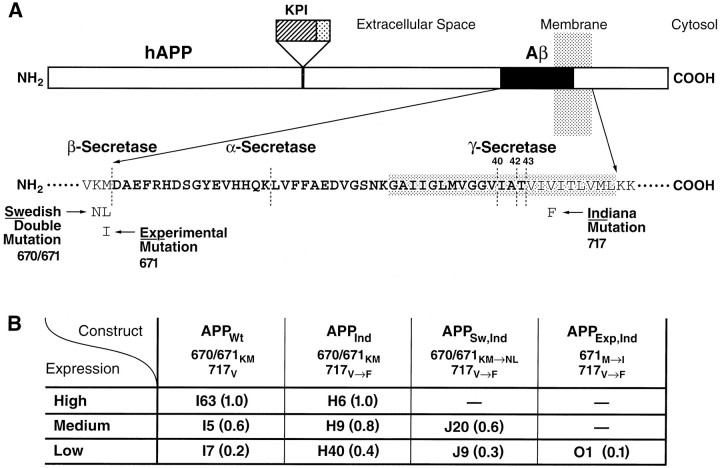
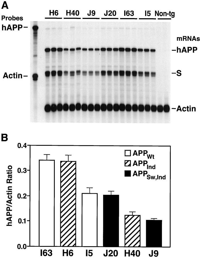
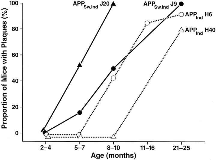
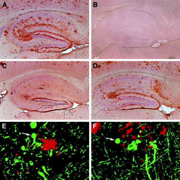
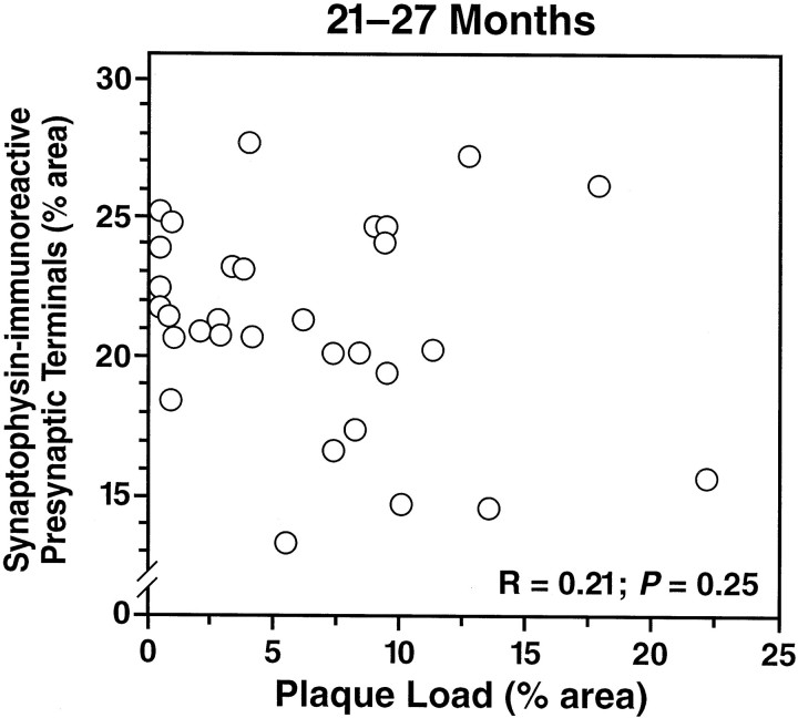
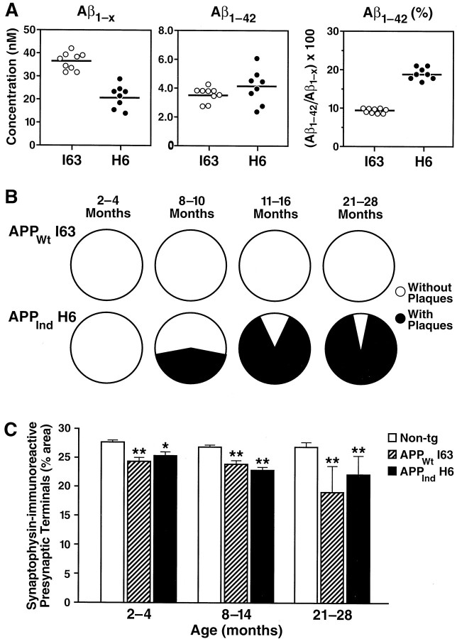
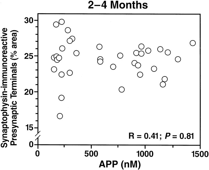
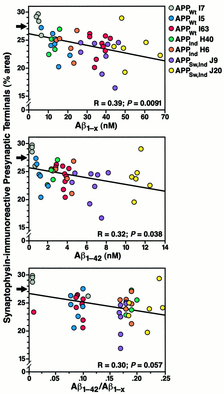
Amyloid plaques are a neuropathological hallmark of Alzheimer's disease (AD), but their relationship to neurodegeneration and dementia remains controversial. In contrast, there is a good correlation in AD between cognitive decline and loss of synaptophysin-immunoreactive (SYN-IR) presynaptic terminals in specific brain regions. We used expression-matched transgenic mouse lines to compare the effects of different human amyloid protein precursors (hAPP) and their products on plaque formation and SYN-IR presynaptic terminals. Four distinct minigenes were generated encoding wild-type hAPP or hAPP carrying mutations that alter the production of amyloidogenic Abeta peptides. The platelet-derived growth factor beta chain promoter was used to express these constructs in neurons. hAPP mutations associated with familial AD (FAD) increased cerebral Abeta(1-42) levels, whereas an experimental mutation of the beta-secretase cleavage site (671(M-->I)) eliminated production of human Abeta. High levels of Abeta(1-42) resulted in age-dependent formation of amyloid plaques in FAD-mutant hAPP mice but not in expression-matched wild-type hAPP mice. Yet, significant decreases in the density of SYN-IR presynaptic terminals were found in both groups of mice. Across mice from different transgenic lines, the density of SYN-IR presynaptic terminals correlated inversely with Abeta levels but not with hAPP levels or plaque load. We conclude that Abeta is synaptotoxic even in the absence of plaques and that high levels of Abeta(1-42) are insufficient to induce plaque formation in mice expressing wild-type hAPP. Our results support the emerging view that plaque-independent Abeta toxicity plays an important role in the development of synaptic deficits in AD and related conditions.


Figures

Fig. 1.
Fig. 1.
Summary of transgenic lines. A, Diagram of hAPP indicating the mutations expressed in transgenic mice. FAD-linked mutations are commonly referred to by place of discovery or residence of affected kindred. The 670/671KM→NL double mutation affects a large pedigree in Sweden (Mullan et al., 1992), and the 717V→F mutation was identified in Indiana (Murrell et al., 1991) (numbers refer to amino acids in APP770). Mutations at position 717 are often collectively referred to as “London mutations” based on the first report of the FAD-linked 717V→I mutation (Goate et al., 1991); however, the latter mutation was not studied here. The sequence of Aβ is indicated inbold in single-letter amino acid code.KPI, Kunitz-type protease inhibitor domain. Elements are not drawn to scale. B, Relative levels of cerebral transgene expression (values in parentheses) were determined in different lines of PDGF-hAPP mice as illustrated in Figure 2. The expression level in line I63 was arbitrarily defined as 1.0.
Fig. 2.
Fig. 2.
Identification of wild-type and FAD-mutant hAPP mice with matching levels of cerebral transgene expression.A, Representative autoradiograph showing results of an RNase protection assay. Total RNA was extracted from entire hemibrains. The left lane shows signals of undigested radiolabeled riboprobes; the other lanes contained the same riboprobes plus brain RNA samples, digested with RNases. Each samplelane contains RNA from a different mouse. The hAPP probe detects human but not mouse APP; it also recognizes an SV40 segment (S) of transgene-derived mRNAs.Non-tg, Nontransgenic. B, Phosphorimager quantitation of signals shown in A. Values represent group means ± SD.
Fig. 3.
Fig. 3.
Comparison of human Aβ levels in hippocampi of mice expressing wild-type or FAD-mutant hAPP. Aβ1-x and Aβ1–42 were quantitated by ELISA in mice from different transgenic lines (n = 6–9 mice per line) at 2–4 months of age. Values represent group means ± SD. No plaques were detected in the opposite hemibrains of these mice by immunostaining with the 3D6 antibody (data not shown).
Fig. 4.
Fig. 4.
Hippocampal plaque formation in different lines of FAD-mutant hAPP mice. Aβ deposits were detected by immunostaining of brain sections (n = 3 per mouse) with the 3D6 antibody as described in Materials and Methods. Six to 18 mice per line were analyzed at 2–4, 8–10, and 21–25 months of age, and 1–6 (mean = 4.3) mice per line were analyzed at 5–7 and 11–16 months of age.
Fig. 5.
Fig. 5.
Age-related cerebral Aβ deposition occurs in mice expressing FAD-mutant hAPP but not in mice expressing wild-type hAPP. Brain sections were immunoperoxidase-stained for Aβ with the 3D6 antibody and imaged by light microscopy (A–D) or double-labeled with antibodies against Aβ (R1280; red) and monoclonal antibodies against phosphorylated neurofilaments (SMI312; green) and imaged by laser scanning confocal microscopy (E,F). Hippocampal sections of transgenic mice are shown: A, APPInd line H6 (18 months);B, APPWt line I63 (15 months);C, APPSw, Ind line J9 (10 months);D, APPSw,Ind line J20 (10 months);E, APPInd line H6 (10 months).F, Midfrontal gyrus from a human AD brain. Magnifications: A–D, 4×; E,F, 930×.
Fig. 6.
Fig. 6.
Density of SYN-IR presynaptic terminals does not correlate with plaque load in mice from different FAD-mutant hAPP lines. Plaque load and density of SYN-IR presynaptic terminals in the hippocampus were determined in 31 transgenic mice from APPInd lines H6, H9, and H40 and APPSw, Indline J9 at 21–27 months of age. No correlation was found between the two variables.
Fig. 7.
Fig. 7.
Comparison of hippocampal Aβ levels, plaque formation, and density of SYN-IR presynaptic terminals in the high expresser lines APPWt I63 and APPInd H6. Note that the cerebral hAPP mRNA levels in these lines are very well matched (Figs. 1, 2). A, Levels of human Aβ were determined at 2–4 months of age in 8–9 mice per line by ELISA.Circles represent values in individual mice;horizontal lines indicate group means. B, Proportion of mice in which 3D6-immunoreactive plaques were identified (black) at the ages indicated (n = 4–18 mice per line and age range). C, The density of SYN-IR presynaptic terminals was determined in 4–41 mice per genotype and age range. Data represent group means ± SD. *p < 0.05, **p < 0.01 versus nontransgenic controls (Tukey–Kramer post hoctest).
Fig. 8.
Fig. 8.
Density of SYN-IR presynaptic terminals does not correlate with hAPP levels across hAPP mice from different lines. Levels of full-length plus α-secreted hAPP and density of SYN-IR presynaptic terminals in the hippocampus were determined in 36 transgenic mice from APPWt lines I5, I7, and I63, APPInd lines H6 and H40, and APPSw, Ind line J9 at 2–4 months of age. No correlation was identified between the two variables. No plaques were detected in the opposite hemibrains of these mice by immunostaining with the 3D6 antibody (data not shown).
Fig. 9.
Fig. 9.
Inverse correlation between density of SYN-IR presynaptic terminals and levels of Aβ. At 2–4 months of age, hippocampal levels of Aβ1-x and Aβ1–42 in one hemibrain were correlated with the hippocampal density of SYN-IR presynaptic terminals in the opposite hemibrain in mice from lines expressing APPWt, APPInd, or APPSw, Ind at different levels (4–9 mice per line). None of these mice had plaques by immunostaining with the 3D6 antibody (data not shown). Arrows indicate the normal density of SYN-IR presynaptic terminals in age-matched nontransgenic controls (mean of 29 mice).

References

    1. Alford MF, Masliah E, Hansen LA, Terry RD. A simple dot-immunobinding assay for quantification of synaptophysin-like immunoreactivity in human brain. J Histochem Cytochem. 1994;42:283–287. - PubMed
    1. Alloul K, Sauriol L, Kennedy W, Laurier C, Tessier G, Novosel S, Contandriopoulos A. Alzheimer's disease: a review of the disease, its epidemiology and economic impact. Arch Gerontol Geriatr. 1998;27:189–221. - PubMed
    1. Bartoo GT, Nochlin D, Chang D, Kim Y, Sumi SM. The mean Aβ load in the hippocampus correlates with duration and severity of dementia in subgroups of Alzheimer disease. J Neuropathol Exp Neurol. 1997;56:531–540. - PubMed
    1. Borchelt DR, Ratovitski T, Van Lare J, Lee MK, Gonzales V, Jenkins NA, Copeland NG, Price DL, Sisodia SS. Accelerated amyloid deposition in the brains of transgenic mice coexpressing mutant presenilin 1 and amyloid precursor proteins. Neuron. 1997;19:939–945. - PubMed
    1. Braak H, Braak E. Evolution of neuronal changes in the course of Alzheimer's disease. J Neural Transm [Suppl 105] 1998;53:127–140. - PubMed

Publication types

MeSH terms

LinkOut - more resources