. 2000 Jun 1;20(11):4069-80.
doi: 10.1523/JNEUROSCI.20-11-04069.2000.

Identification of proteins in the postsynaptic density fraction by mass spectrometry

Affiliations

Identification of proteins in the postsynaptic density fraction by mass spectrometry

R S Walikonis et al. J Neurosci. .

Abstract

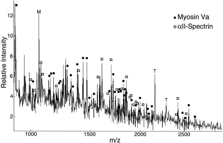
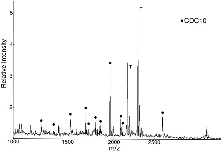
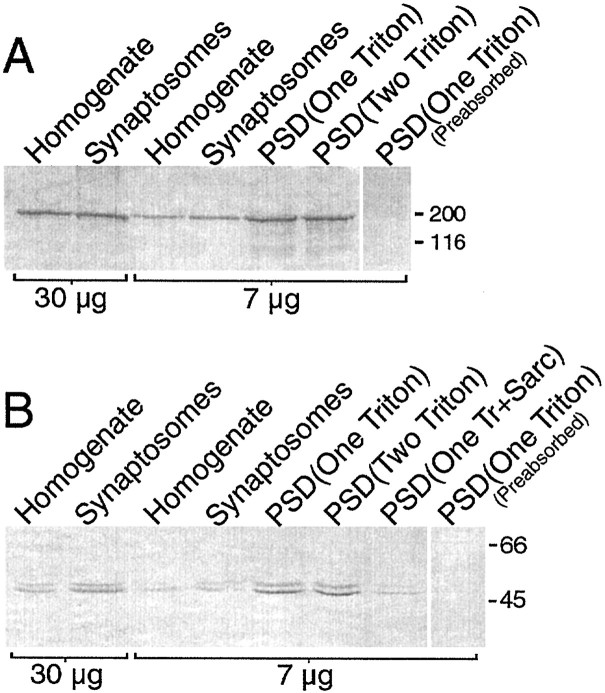
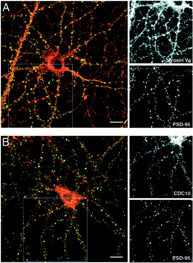
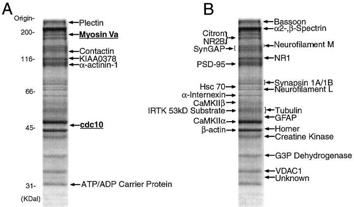
Our understanding of the organization of postsynaptic signaling systems at excitatory synapses has been aided by the identification of proteins in the postsynaptic density (PSD) fraction, a subcellular fraction enriched in structures with the morphology of PSDs. In this study, we have completed the identification of most major proteins in the PSD fraction with the use of an analytical method based on mass spectrometry coupled with searching of the protein sequence databases. At least one protein in each of 26 prominent protein bands from the PSD fraction has now been identified. We found 7 proteins not previously known to be constituents of the PSD fraction and 24 that had previously been associated with the PSD by other methods. The newly identified proteins include the heavy chain of myosin-Va (dilute myosin), a motor protein thought to be involved in vesicle trafficking, and the mammalian homolog of the yeast septin protein cdc10, which is important for bud formation in yeast. Both myosin-Va and cdc10 are threefold to fivefold enriched in the PSD fraction over brain homogenates. Immunocytochemical localization of myosin-Va in cultured hippocampal neurons shows that it partially colocalizes with PSD-95 at synapses and is also diffusely localized in cell bodies, dendrites, and axons. Cdc10 has a punctate distribution in cell bodies and dendrites, with some of the puncta colocalizing with PSD-95. The results support a role for myosin-Va in transport of materials into spines and for septins in the formation or maintenance of spines.


Figures

Fig. 1.
Fig. 1.
Strategy for identifying proteins in the PSD fraction by MALDI-TOF mass spectrometry. The masses of tryptic peptides derived from individual protein bands in an SDS-PAGE gel are compared with theoretical masses of tryptic peptides for each protein in the database.
Fig. 2.
Fig. 2.
MALDI-TOF peptide mass map obtained from a 190 kDa protein band in the PSD fraction. Ion signals with measured masses that match calculated masses of protonated tryptic peptides of myosin-Va (●) and αII-spectrin (■) within 50 ppm are indicated. T, Signals from autolysis products of trypsin; M, signals from matrix-related ions.
Fig. 3.
Fig. 3.
MALDI-TOF peptide mass map obtained from a 45 kDa protein band in the PSD fraction. Ion signals with measured masses that match calculated masses of mouse cdc10 (●) within 50 ppm are indicated. T, Signals derived from autolysis products of trypsin.
Fig. 4.
Fig. 4.
Myosin-Va and cdc10 are enriched in the PSD fraction. Immunoblots were prepared with 30 μg (lanes 1, 2) and 7 μg (lanes 3–6) each of rat forebrain homogenate, synaptosome fraction, One-Triton fraction, and Two-Triton fraction, prepared as described in Materials and Methods. B also includes 7 μg of a One-Triton plus Sarcosyl PSD fraction in lane 7. Blots prepared with antibodies preabsorbed with their respective antigens are shown in the right lane.A, The myosin-Va protein bands were visualized with antibody DB1C at 1:1500 dilution. B, The cdc10 protein bands were visualized with antibody L2 at 1:1000 dilution.
Fig. 5.
Fig. 5.
Immunocytochemical localization of myosin-Va and cdc10 in cultures of dissociated hippocampal neurons. Hippocampal neurons dissociated at E18 were grown in culture for 28 d and then fixed and double-immunostained as described in Materials and Methods. Images of the two fluorophors were colorized and combined (left). At right are the single images of the boxed regions. A, Immunocytochemical localization of myosin-Va and PSD-95.Red indicates Cy3 staining of myosin V, andgreen indicates FITC staining of PSD-95. Regions of overlap are yellow. Myosin-Va is distributed throughout cell bodies, dendrites, and axons and appears concentrated at synapses, where it colocalizes with PSD-95. B, Immunocytochemical localization of cdc10 and PSD-95. Red indicates Cy3 staining of cdc10, and green indicates FITC staining of PSD-95. CDC10 has a punctate distribution throughout the soma and dendrites. Some, but not all, of the cdc10 in dendrites colocalizes with PSD-95 at synapses. Scale bars, 10 μm.
Fig. 6.
Fig. 6.
Proteins identified in the PSD fraction by MALDI-TOF MS. Thirty micrograms of protein from the Two-Triton PSD fraction were subjected to SDS-PAGE and stained with Coomassie blue. Individual protein bands were isolated, and the proteins in each band were identified by MALDI-TOF mass spectrometry as described in Materials and Methods. The positions of molecular weight standards are shown at left. A, Proteins identified for the first time in this study as constituents of the PSD fraction. The presence of myosin-Va and cdc10 (bold,underlined) at synaptic sites was verified in this study. B, Proteins identified in this study that were previously identified in the PSD fraction by other methods.

References

    1. Abbott M-A, Wells DG, Fallon JR. The insulin receptor tyrosine kinase substrate p58/53 and the insulin receptor are components of CNS synapses. J Neurosci. 1999;19:7300–7308. - PMC - PubMed
    1. Adam G, Matus A. Role of actin in the organization of brain postsynaptic densities. Brain Res Mol Brain Res. 1996;43:246–250. - PubMed
    1. Apperson ML, Moon I-S, Kennedy MB. Characterization of densin-180, a new brain-specific synaptic protein of the O-sialoglycoprotein family. J Neurosci. 1996;16:6839–6852. - PMC - PubMed
    1. Benfenati F, Valtorta F, Rubenstein JL, Gorelick FS, Greengard P, Czernik AJ. Synaptic vesicle-associated Ca2+/calmodulin-dependent protein kinase II is a binding-protein for synapsin I. Nature. 1992;359:417–420. - PubMed
    1. Bessman SP, Carpenter CL. The creatine-creatine phosphate energy shuttle. Annu Rev Biochem. 1985;54:831–862. - PubMed

Publication types

MeSH terms

LinkOut - more resources