. 2000 Jun 1;20(11):4081-90.
doi: 10.1523/JNEUROSCI.20-11-04081.2000.

Ciliary neurotrophic factor and stress stimuli activate the Jak-STAT pathway in retinal neurons and glia

Affiliations

Ciliary neurotrophic factor and stress stimuli activate the Jak-STAT pathway in retinal neurons and glia

W M Peterson et al. J Neurosci. .

Abstract

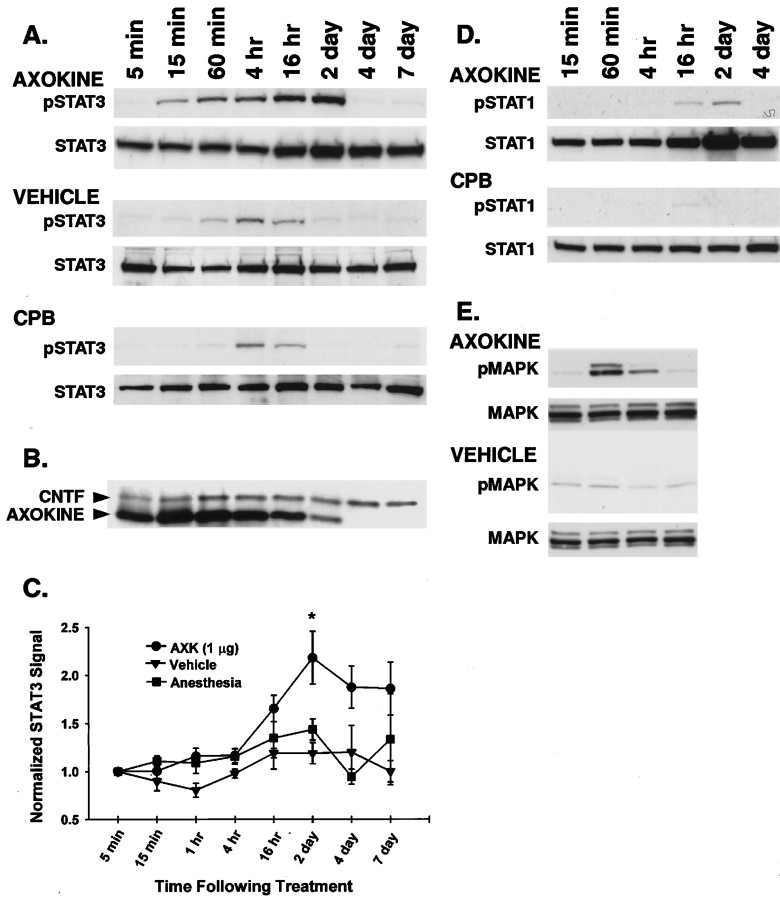
Ciliary neurotrophic factor (CNTF) is pleiotrophic for central, peripheral, and sensory neurons. In the mature retina, CNTF treatment enhances survival of retinal ganglion and photoreceptor cells exposed to otherwise lethal perturbation. To understand its mechanism of action in vivo, the adult rat retina was used as a model to investigate CNTF-mediated activation of Janus kinase/signal transducer and activator of transcription (Jak-STAT) and ras-mitogen activated protein kinase (ras-MAPK). Intravitreal injection of Axokine, an analog of CNTF, phosphorylates STAT3 and MAPK and produces delayed upregulation of total STAT3 and STAT1 protein in rat retina. Activated STAT3 is predominantly localized in nuclei of retinal Müller (glial) cells, ganglion cells, and astrocytes, but not in photoreceptors. Although CNTF alpha-receptor (CNTFRalpha) mRNA and protein are localized predominantly if not exclusively in retinal neurons, coincident CNTF-mediated STAT3 signaling was observed in both glia and neurons. CNTF-induced activation of Jak-STAT signaling prompted us to investigate STAT3 phosphorylation after a variety of stress-mediated, conditioning stimuli. We show that STAT3 is activated in the retina after exposure to subtoxic bright light, mechanical trauma, and systemic administration of the alpha(2)-adrenergic agonist xylazine, all of which have been shown previously to condition photoreceptors to resist light-induced degeneration. These results demonstrate that CNTF directly stimulates Jak-STAT and ras-MAPK cascades in vivo and strongly suggest that STAT3 signaling is an underlying component of neural responsiveness to stress stimuli. The observation that CNTF activates STAT3 in ganglion cells, but not in photoreceptors, suggests that Jak-STAT signaling influences neuronal survival via both direct and indirect modes of action.


Figures

Fig. 1.
Fig. 1.
Effects of Axokine on phosphorylated and total STAT3, STAT1, and MAPK in homogenized rat retinas. A, Retinas were harvested at various times indicated (5 min to 7 d) after a single intravitreal injection of Axokine. Homogenized retinas were Western blotted (50 μg/lane) to detect total STAT3 and Y705-phosphorylated STAT3 (pSTAT3). The band size for both STAT3 and pSTAT3 is the expected 92 kDa. Axokine produced robust pSTAT3 signal beginning at 15 min and disappearing by 4 d. Weaker and more transient pSTAT3 signals were seen in the vehicle and anesthesia (CPB) control groups. B, Membranes from Axokine-treated STAT3/pSTAT3 immunoblots were stripped and reblotted using an antibody that recognizes both CNTF and Axokine (23 and 21 kDa, respectively). Axokine was detected in the retina for up to 2 d after injection. C, Graph summarizing the time-dependent effects of total STAT3 for each treatment group. STAT3 signal was quantified at each time point using conventional densitometric analysis and normalized to the STAT3 signal at 5 min. Analysis of immunoblots from time course studies culled from three separate groups of animals shows a significant increase in normalized STAT3 signal in the Axokine-treated group (p < 0.05; repeated ANOVA, Dunnett's post hocttest). Error bars are in terms of SE. D, Immunoblots for total STAT1 and Y701-STAT1 (pSTAT1) were performed on Axokine-treated and anesthesia control retinas. Axokine weakly activated pSTAT1 at the later time points but dramatically increased total STAT1 levels by 16 hr. E, Western blots for total MAPK and pMAPK for Axokine-treated retinas showed robust transient activation of MAPK at 60 min.
Fig. 2.
Fig. 2.
Immunolocalization of total STAT3 in vehicle- and Axokine-treated retinal cross-sections viewed with differential interference contrast optics. A, Injection of vehicle (VEH) buffer 15 min before the animals were killed exhibited the same STAT3 labeling pattern as untreated eyes (latter not shown). In the inner retina, diffuse labeling of STAT3 was seen along the NFL and GCL, and along radial processes within the IPL and INL (thin arrow). Weak STAT3 immunoreactivity was apparent in the OPL but not in the photoreceptor layer.B, Within 15 min of Axokine (AXK) injection, we observed intense labeling of nuclei along the NFL (thick arrow), heavier labeling of GC bodies and nuclei (arrowhead), and heavier labeling of cell bodies and nuclei in the INL (thin arrow) near the injection site.C, No specific labeling was observed in the absence of primary antibody. D, Retinal cross-section taken from four contiguous regions, extending from near the injection site (right half of panel) to further away (left half of panel).E–H, Magnification of boxed regions ofD showing spatial profile of STAT3 labeling from regions distal to the injection site (E) to regions closest to the injection site (H). Proximity to the injection site is associated with a progressive increase in STAT3 labeling of cell bodies and nuclei in INL and a concomitant gradual decrease of labeling in processes in the IPL, suggesting mobilization of STAT3 from cytoplasm to cell body, and then to nucleus. Heavy labeling of nuclei in the NFL and GCL is seen throughout the entire region. RPE, Retinal pigment epithelium; OS, photoreceptor outer segments; IS, photoreceptor inner segments.
Fig. 3.
Fig. 3.
Effects of Axokine on pSTAT3 immunolocalization and colocalization of pSTAT3 and S-100 in Müller cells.A, In 15 min vehicle-treated (VEH) eyes, sparse pSTAT3 labeling is present only along the NFL.B, Labeling of astrocytes and GC nuclei is observed adjacent to the injection site 15 min after Axokine (AXK) treatment. C,D, Two representative regions of pSTAT3 immunoreactivity in the same retina 30 min after Axokine treatment. C, pSTAT3 heavily labeled GC nuclei and weakly labeled nuclei in the INL.D, pSTAT3 heavily labeled nuclei in the INL but produced little labeling in GC bodies and nuclei. E, At 1 hr, strong labeling of nuclei in INL (thin arrow), GCs (thick arrow), and astrocytes (arrowhead) was observed in most of the retina. F, A diffuse band of pSTAT3 labeling was evident along the GCL and NFL in vehicle-treated eyes. G, pSTAT3 labeling of nuclei in the INL gradually disappeared by 4 hr after Axokine treatment, but signal could still be detected in nuclei within the GCL. H, At 16 hr after Axokine injection, no specific pSTAT3 immunoreactivity was detected. I, J, One-to-one colocalization of S-100 antigen (left) and pSTAT3 (right) immunoreactivity in the INL is consistent with exclusive localization of STAT3 in Müller cells in the INL.K, At 60 min after Axokine treatment, pSTAT3 immunoreactivity was observed in nuclei of astrocytes throughout the optic nerve head (ONH). RPE, Retinal pigment epithelium; OS, photoreceptor outer segments; IS, photoreceptor inner segments.
Fig. 4.
Fig. 4.
Immunolocalization of pMAPK. A, At 30 min after Axokine (AXK) injection, intense pMAPK staining is noted in cells and cell processes in the NFL, particularly surrounding blood vessels, which is consistent with staining of astrocytes. Intense staining of nuclei in the INL and of radial elements extending from the NFL through the ONL is consistent with distribution of pMAPK throughout Müller cell bodies and processes. At this time point, pMAPK staining was strongest in regions nearest the injection site. B, Significantly weaker pMAPK staining along the NFL in vehicle-treated (VEH) eyes in regions proximal to the injection site suggests stress-induced activation of MAPK in astrocytes. Very little staining of pMAPK was observed in Müller cells.C, Representative photomicrograph of the uniform pMAPK staining apparent within the entire retina at 60 min after Axokine injection. Note the appearance of pMAPK labeling along the outer limiting membrane. D, Very little labeling of pMAPK was observed in 1 hr vehicle-treated eyes.
Fig. 5.
Fig. 5.
Time course of Axokine-induced expression of GFAP in Müller cells. At 16 hr, vehicle-injected (VEH) and Axokine-injected (AXK) retinas still showed essentially normal GFAP labeling, restricted to astrocyte processes and end feet of Müller cells along the NFL and surrounding blood vessels. At 2 d after Axokine injection, GFAP labeled radial processes in the IPL. At 7 d, labeling of processes throughout the IPL and into the ONL was observed in Axokine-treated eyes but not vehicle-treated eyes. Labeling of radial processes throughout the IPL persisted for at least 3 weeks after Axokine treatment.
Fig. 6.
Fig. 6.
Activation of STAT3 and/or STAT1 after conditioning with 24 or 48 hr of bright light or treatment with various combinations of ketamine and xylazine. In A, animals were recovered in normal lighting conditions for the time periods shown before retinas were harvested for immunoblotting. A, STAT3 was activated immediately after 24 or 48 hr of constant bright light (day 0). An increase in total STAT3 levels was also observed in both conditioning groups. Twenty-four and 48 hr of bright light produced a small, delayed increase in pSTAT1 signal during the recovery period and a robust increase in total STAT1 levels. B, Effects of various combinations of ketamine and xylazine (intraperitoneal injection) on pSTAT3 and total STAT3 signal in retina. Xylazine alone or in combination with ketamine produced a robust phosphorylation of STAT3 4 hr after administration. No activation of STAT3 was observed in the ketamine group.
Fig. 7.
Fig. 7.
Needle injury to the retina induces GFAP upregulation and increases total STAT3 labeling around the injury site.A, Fluorescent photomicrograph of robust GFAP-positive processes in the GCL (which borders the inner limiting membrane) and throughout the retina encompassing the region of trauma.B, GFAP labeling in the same eye in a region of the retina that is far from the site of injury. Normal GFAP-positive labeling in the region between the GCL and the ILM is observed.C, In a representative section from the same region asA, increased STAT3 signal is detected along ILM, the GCL, and radial processes in the IPL, as well as cell bodies and nuclei in the INL. D, In a representative section from the same region as B, STAT3 immunoreactivity in regions far from the injection site is similar to that of unperturbed retinas.RPE, Retinal pigment epithelium; OS, photoreceptor outer segments.

References

    1. Acarin L, Gonzalez B, Castellano B. Stat3 and NFκB glial expression after excitotoxic damage to the postnatal brain. NeuroReport. 1998;9:2869–2873. - PubMed
    1. Adler R, Landa KB, Manthorpe M, Varon S. Cholinergic neuronotrophic factors: intraocular distribution of trophic activity for ciliary neurons. Science. 1979;204:1434–1436. - PubMed
    1. Anderson KD, Panayotatos N, Corcoran TL, Lindsay RM, Wiegand SJ. Ciliary neurotrophic factor protects striatal output neurons in an animal model of Huntington disease. Proc Natl Acad Sci USA. 1996;93:7346–7351. - PMC - PubMed
    1. Bonni A, Frank DA, Schindler C, Greenberg ME. Characterization of a pathway for ciliary neurotrophic factor signaling to the nucleus. Science. 1993;262:1575–1579. - PubMed
    1. Bonni A, Sun Y, Nadal-Vicens M, Bhatt A, Frank DA, Rozovsky I, Stahl N, Yancopoulos GD, Greenberg ME. Regulation of gliogenesis in the central nervous system by the JAK-STAT signaling pathway. Science. 1997;278:477–483. - PubMed

MeSH terms

Substances