. 2000 Jun 1;20(11):4145-55.
doi: 10.1523/JNEUROSCI.20-11-04145.2000.

A-type K+ current mediated by the Kv4 channel regulates the generation of action potential in developing cerebellar granule cells

Affiliations

A-type K+ current mediated by the Kv4 channel regulates the generation of action potential in developing cerebellar granule cells

R Shibata et al. J Neurosci. .

Abstract

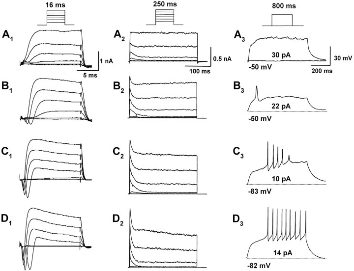
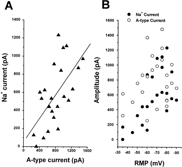
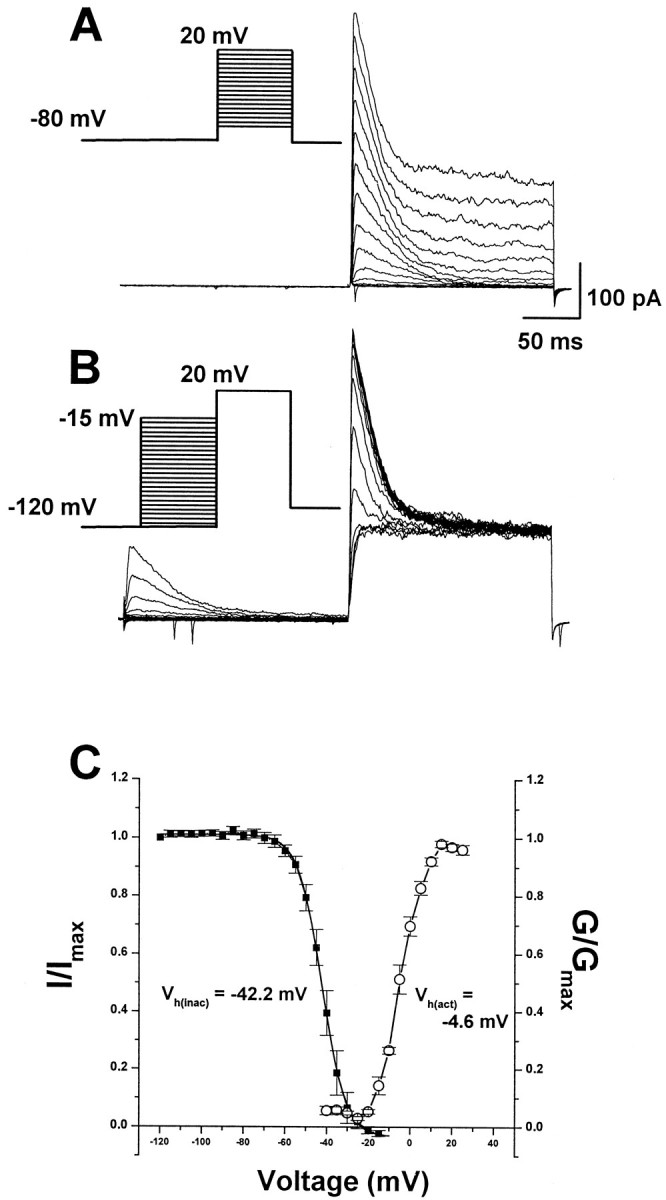
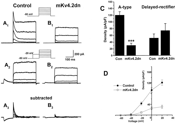
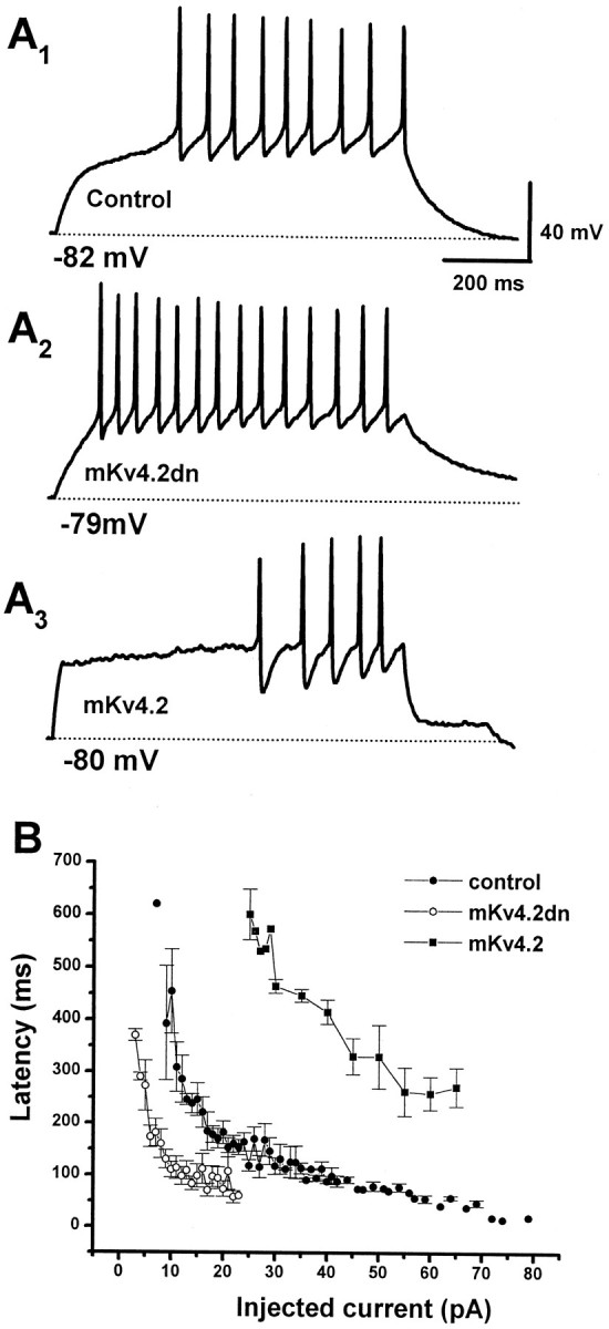
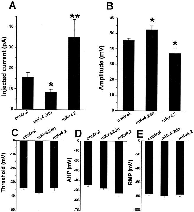
During neuronal differentiation and maturation, electrical excitability is essential for proper gene expression and the formation of synapses. The expression of ion channels is crucial for this process; in particular, voltage-gated K(+) channels function as the key determinants of membrane excitability. Previously, we reported that the A-type K(+) current (I(A)) and Kv4.2 K(+) channel subunit expression increased in cultured cerebellar granule cells with time. To examine the correlation between ion currents and the action potential, in the present study, we measured developmental changes of action potentials in cultured granule cells using the whole-cell patch-clamp method. In addition to an observed increment of I(A), we found that the Na(+) current also increased during development. The increase in both currents was accompanied by a change in the membrane excitability from the nonspiking type to the repetitive firing type. Next, to elucidate whether Kv4.2 is responsible for the I(A) and to assess the effect of Kv4 subunits on action potential waveform, we transfected a cDNA encoding a dominant-negative mutant Kv4.2 (Kv4.2dn) into cultured cells. Expression of Kv4.2dn resulted in the elimination of I(A) in the granule cells. This result demonstrates that members of the Kv4 subfamily are responsible for the I(A) in developing granule cells. Moreover, elimination of I(A) resulted in shortening of latency before the first spike generation. In contrast, expression of wild-type Kv4.2 resulted in a delay in latency. This indicates that appearance of I(A) is critically required for suppression of the excitability of granule cells during their maturation.


Figures

Fig. 1.
Fig. 1.
Expression of EGFP in microexplant cultures using the lipofection method. A, A lower magnification of EGFP-positive cells near an explant at 7 DIV. The border of the explant is indicated by a line. The EGFP expression vector was transfected at 4 DIV. Approximately 10 cells per explant were labeled with EGFP. Most positive cells had short dendrites and a pair of long processes extending radially to the explant. These cells were typical granule cells. B, Representative bipolar cell observed at 2 DIV. EGFP cDNA was transfected at 1 DIV.C, Typical T-shaped cell observed at 7 DIV. Thearrow points to the T-junction. D, Many mature granule cells had dendrites but did not exhibit T-shape. Scale bars: A, 50 μm; B–D, 10 μm.
Fig. 2.
Fig. 2.
Developmental changes in membrane current and action potential in the granule cells. A, Representative whole-cell current (A1 andA2) and voltage response (A3) recorded from a bipolar cell at 2 DIV. Records were obtained from the same cell.A1, Cell was elicited with step depolarizations from a holding potential of −80 mV to a potential of between −60 and +40 mV with 20 mV increments for 16 msec in voltage-clamp mode to monitor fast inward currents (inset; the same protocol was applied inB1,C1, andD1). Fast-rising inward currents were not evoked in bipolar cells. A2, The same protocol as shown in A1 was applied for 250 msec to monitor outward currents (this protocol was also applied inB1,C1, andD1).A3, A depolarizing current step of 30 pA for 800 msec did not evoke action potential from a holding potential of −50 mV. B–D, Representative whole-cell currents (B1,B2,C1,C2,D1, andD2) and voltage responses (B3,C3, andD3) recorded from T-shaped cells at 4–7 DIV. Recordings in B, C, andD were obtained from the same cell. Fast-rising inward currents and fast-inactivating currents increased in T-shaped cells.B3,C3,D3, A depolarizing current step of 22 (B3), 10 (C3), or 14 (D3) pA was applied for 800 msec from a holding potential of −50 (B3), −83 (C3), or −82 (D3) mV, respectively. In the T-shaped cells, three types of discharge patterns, single spike (B3), rapidly adapting (C3), and repetitive firing (D3), were observed.
Fig. 3.
Fig. 3.
A-Type currents and Na+currents increased over a similar time course. A, The amplitude Na+ current was plotted as a function of A-type current recorded from the same cell. These currents were recorded from a mix of bipolar and T-shaped cells. A-Type current was isolated by the prepulse protocol, and its peak amplitude was measured. The relationship between the A-type current and the Na+ current was fitted by the least-squares method (solid line; r = 0.65).B, The size of the Na+ current (filled circles) and the A-type current (open circles) was plotted as a function of RMP. Note that the increase in amplitude of both currents correlated with the depth of the RMP.
Fig. 4.
Fig. 4.
Voltage dependence of activation and inactivation of transient current in the granule cells. A, Superimposed current traces evoked by depolarizing steps to potentials between −60 and +20 mV with 5 mV increment after −80 mV.B, Superimposed current traces evoked by test depolarization to +20 mV after 200 msec prepulse to potentials between −120 and −15 mV with 5 mV increment. C, Plot of normalized peak current as a function of conditioning voltage. Boltzmann functions with half-activation voltage of −4.6 mV and half-inactivation voltage of −42.2 mV. Spontaneous inward spikes occasionally remained after blockade of spontaneous activity by TTX and Ca2+ channel blocker.
Fig. 5.
Fig. 5.
Recovery from inactivation of A-type currents.A, Inactivation recovery was examined by inactivating the A-type current and then stepping to −120, −80, or −50 mV for increasing before a test step to 20 mV. The voltage protocol is shown above the current traces. Current traces recovered from −120 (top traces), −80 (middle traces), and −50 (bottom traces) mV are shown. B, Plots of peak current as a function of prepulse duration at −120 (filled circles), −80 (open circles), and −50 (filled squares) mV. Data were fitted with a single exponential with time constants of 6.6 (at −120 mV), 15.5 (at −80 mV), and 41.4 (at −50 mV) msec, respectively.
Fig. 6.
Fig. 6.
mKv4.2dn suppressed the mKv4.2 current in transiently transfected HEK293 cells.A1, Wild-type mKv4.2 current was obtained when mKv4.2 and pCR3.1 vectors were cotransfected into HEK293 cells. Cells were held at −80 mV and then stepped to test potentials ranging from −60 to +40 mV (in 20 mV increments) for 250 msec.A2, Wild-type mKv4.2 current was functionally eliminated when mKv4.2 and Kv4.2dn were cotransfected. Currents were evoked as described in A1.B, Mean amplitude of peak currents at a +40 mV test pulse in HEK293 cells expressed with mKv4.2, mKv4.2 plus mKv4.2dn, mKv4.2dn, and pCR3.1E (mean ± SEM). The amplitude of the endogenous current expressed in HEK293 cells was measured from cells transfected with pCR3.1E. mKv4.2dn did not produce a functional current. The differences between mKv4.2 and mKv4.2 plus mKv4.2dn are statistically significant (Student's t test; ***p < 0.001). C, D, Kv1.1 current (C1) or Kv3.1 current (D1) was obtained when Kv3.1 were cotransfected into CHO-K1 cells. Cells were held at −80 mV and then stepped to test potentials ranging from −60 to +40 mV (in 20 mV increments) for 250 msec. Neither Kv1.1 current (C2) nor Kv3.1 current (D2) was suppressed by cotransfection with mKv4.2dn. E, Mean amplitude of peak currents at a +40 mV test pulse in CHO-K1 cells expressed with Kv1.1, Kv1.1 plus mKv4.2dn, Kv3.1, and Kv3.1 plus mKv4.2dn (mean ± SEM).
Fig. 7.
Fig. 7.
Expression of mKv4.2dn suppressed the A-type current of cerebellar granule cells.A1, Cerebellar granule cells transfected with EGFP in the microexplant culture exhibited a large transient and maintained outward current by a series of depolarizing pulses of −60 to +40 mV. A2, The transient component of the current can be inactivated with a prepulse to −20 mV. A3, Isolated A-type current was obtained by subtracting A2 fromA1.B1B3, Cotransfection of mKv4.2dn and EGFP results in a marked suppression of the transient component without affecting the maintained component of outward currents. C, Quantitative analysis indicated the suppression of A-type current and no effect on delayed rectifier current in the peak density evoked at 20 mV. Mean ± SEM is displayed. ***p < 0.001 versus control cells.D, Voltage–current density relationship for A-type currents recorded from control cells (filled circles) and mKv4.2dn-transfected cells (open circles). Mean ± SEM is displayed.
Fig. 8.
Fig. 8.
Expression of mKv4.2 altered the inactivation time constant of A-type currents in the cerebellar granule cells.A, Representative current recorded from a granule cell transfected with EGFP alone (A1) and with EGFP plus mKv4.2 (A2). Because the transfection of 0.2 μg/well mKv4.2 DNA caused serious damage to the granule cells (see Discussion), the amount of DNA for transfection was reduced to 0.05 μg/well. A depolarizing pulse to +20 mV from a holding potential of −80 mV was given. B, Time constant of inactivation as a function of voltage in control cells (filled circles) and mKv4.2-transfected cells (open circles). Time constant of inactivation of the mKv4.2 current in HEK293 cells (filled triangles) is also plotted. Mean ± SEM is displayed. C, Voltage–current density relationship for A-type currents recorded from control cells (filled circles) and mKv4.2-transfected cells (open circles). Mean ± SEM is displayed.
Fig. 9.
Fig. 9.
Effect of elimination or prolonged inactivation kinetics of A-type current on the latency to the first spike.A, Discharge pattern of EGPF-transfected cells (A1, Control), mKv4.2dn plus EGFP-transfected cells (A2), and mKv4.2 plus EGFP-transfected cells (A3). The cells were held at or near −80 mV and injected with a current of 14 (A1), 10 (A2), or 30 (A3) pA for 800 msec.B, The latency to the first spike was plotted as a function of amplitude of injected currents (mean ± SEM;n = 6 for control cells, n = 4 for mKv4.2dn-transfected cells, and n = 5 for mKv4.2-transfected cells). The FSL recorded from mKv4.2dn-transfected cells is shorter and the FSL from mKv4.2-transfected cells is longer compared with the FSL from control cells.
Fig. 10.
Fig. 10.
Effects of mKv4.2dn or mKv4.2dn expression on the physiology of granule cells. A, Minimum amplitude of injected current required for generating spikes. The data indicate that the minimum amplitude of injection was reduced in mKv4.2dn-transfected cells and increased in mKv4.2-transfected cells compared with control cells. Mean ± SEM. The differences between control cells (n = 12) and mKv4.2dn-transfected cells (n = 9, *p < 0.05), or between control cells and mKv4.2-transfected cells (n = 5, **p < 0.01) were statistically significant.B, The amplitude of the first spike. The amplitude was larger in mKv4.2dn-transfected cells and smaller in mKv4.2-transfected cells compared with control cells. Mean ± SEM. The differences between control cells and mKv4.2dn-transfected cells, or between control cells and mKv4.2-transfected cells were statistically significant (*p < 0.05). C–E, The threshold of action potential (C), amplitude of AHP (D), and RMP (E) did not appear to be affected by changes in A-type current properties.

Similar articles

Cited by

References

    1. Arhem P, Johansson S. A model for the fast 4-aminopyridine effects on amphibian myelinated nerve fibres. A study based on voltage-clamp experiments. Acta Physiol Scand. 1989;137:53–61. - PubMed
    1. Baldwin TJ, Tsaur ML, Lopez GA, Jan YN, Jan LY. Characterization of a mammalian cDNA for an inactivating voltage-sensitive K+ channel. Neuron. 1991;7:471–483. - PubMed
    1. Bardoni R, Belluzzi O. Kinetic study and numerical reconstruction of A-type current in granule cells of rat cerebellar slices. J Neurophysiol. 1993;69:2222–2231. - PubMed
    1. Belluzzi O, Sacchi O, Wanke E. A fast transient outward current in the rat sympathetic neurone studied under voltage-clamp conditions. J Physiol (Lond) 1985;358:91–108. - PMC - PubMed
    1. Campbell DL, Qu Y, Rasmusson RL, Strauss HC. The calcium-independent transient outward potassium current in isolated ferret right ventricular myocytes. II. Closed state reverse use-dependent block by 4-aminopyridine. J Gen Physiol. 1993;101:603–626. - PMC - PubMed

Publication types

MeSH terms