. 2000 Jun 1;20(11):4255-66.
doi: 10.1523/JNEUROSCI.20-11-04255.2000.

Evidence that separate neural circuits in the nucleus accumbens encode cocaine versus "natural" (water and food) reward

Affiliations

Evidence that separate neural circuits in the nucleus accumbens encode cocaine versus "natural" (water and food) reward

R M Carelli et al. J Neurosci. .

Abstract

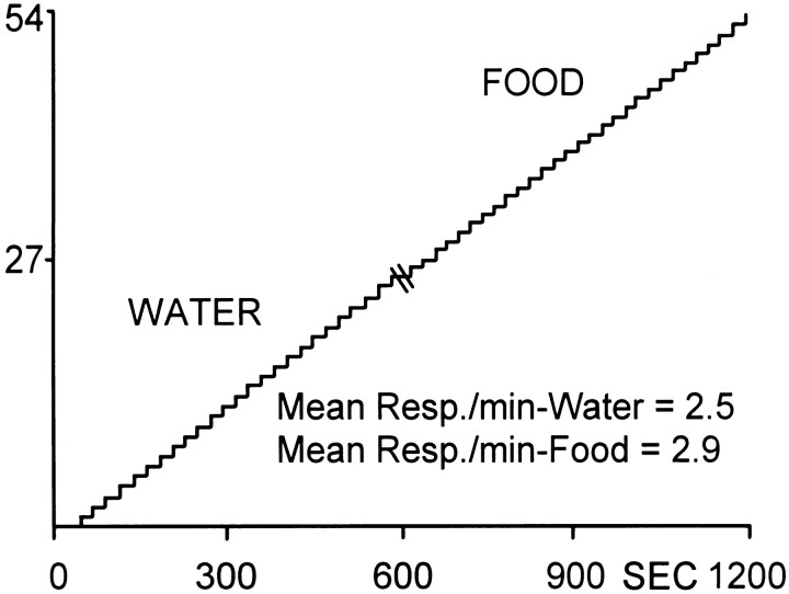
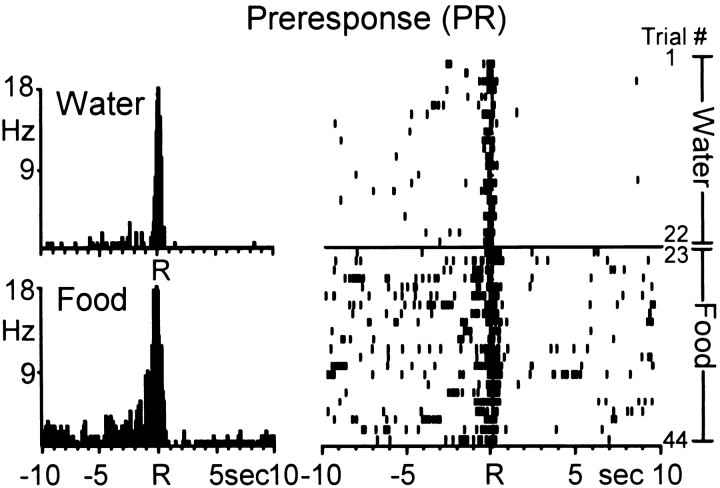
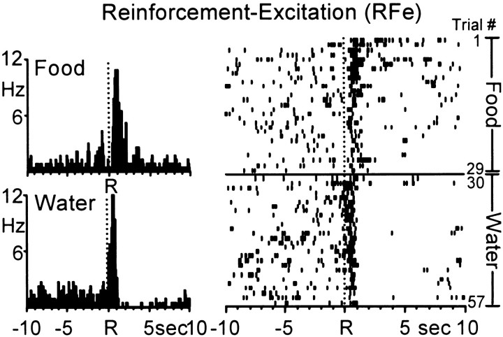
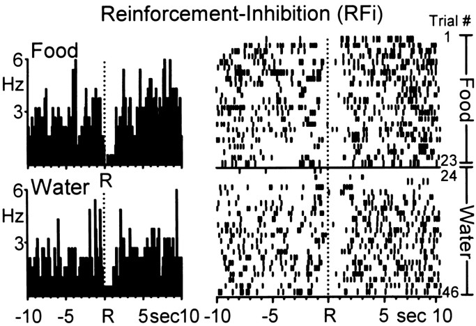
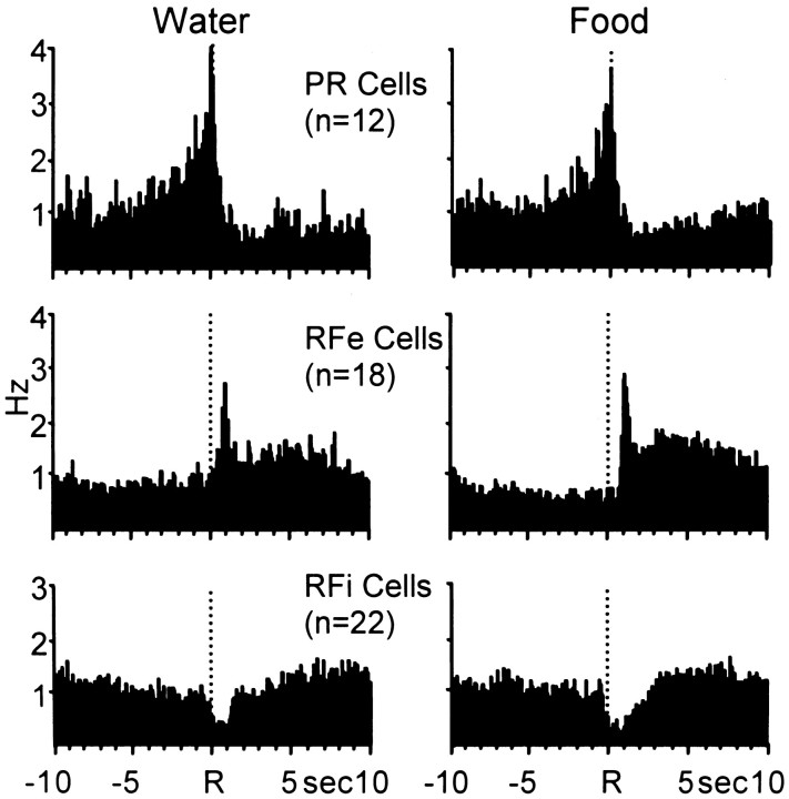
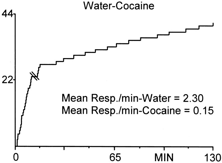
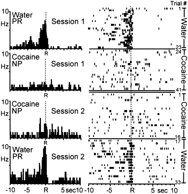
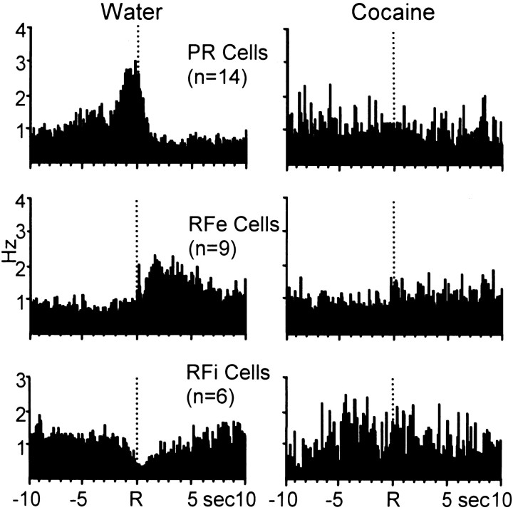
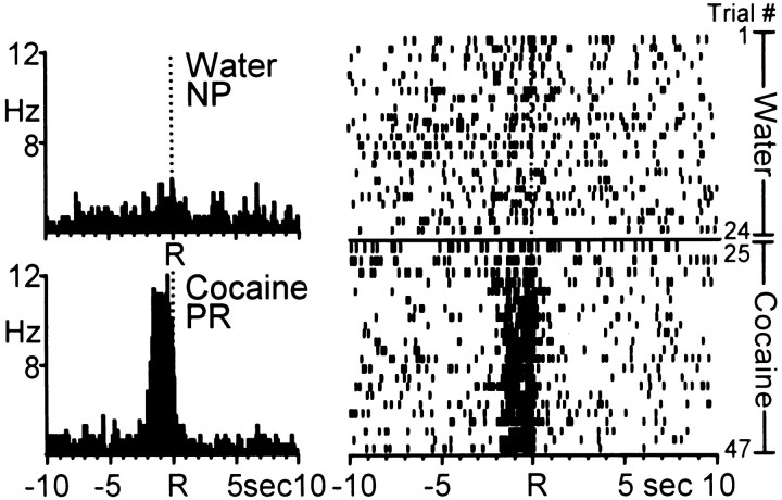
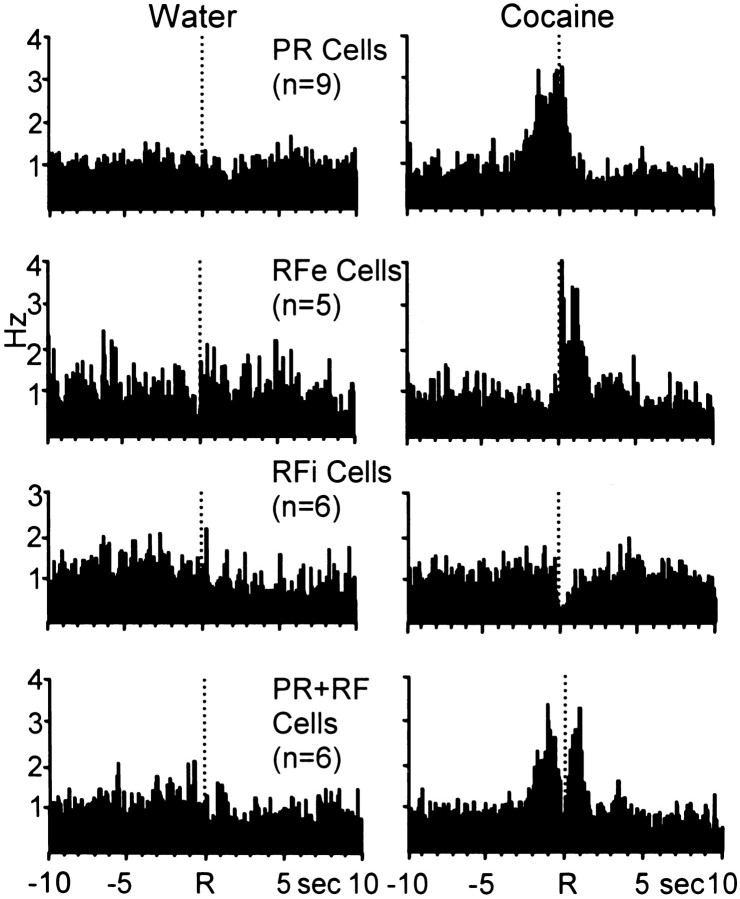
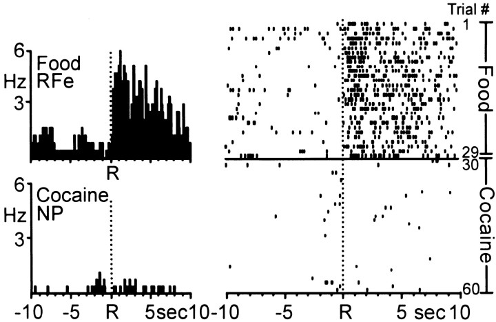
Electrophysiological recording procedures were used to examine nucleus accumbens (Acb) cell firing in rats trained to press a lever on a multiple schedule [ fixed ratio (FR)1, FR1] for either two "natural" reinforcers (food and water), or a natural reinforcer and intravenous self-administration of cocaine. Of 180 cells recorded during water and food reinforcement (n = 13 rats), 77 neurons were classified as phasically active, exhibiting one of three well-defined types of patterned discharges relative to the reinforced-response (Carelli and Deadwyler, 1994). Of the 77 phasic cells, the majority (68%) showed similar types of patterned discharges across the two natural reinforcer conditions. In contrast, of 127 neurons recorded during water and cocaine reinforcement (n = 8 rats), only 5 of 60 phasically active cells (8%) exhibited similar types of patterned discharges relative to water- and cocaine-reinforced responding. The remaining 55 phasic cells (92%) displayed patterned discharges relative to the cocaine-reinforced response (n = 26 cells), or relative to the water-reinforced response (n = 29 cells), but not both. For some rats (n = 3), food was substituted for water in the task. Again, the majority of phasic neurons (13 of 14 cells, 93%) exhibited nonoverlapping firing patterns across the drug and natural reinforcer conditions. These findings indicate that in the well-trained animal, cocaine activates a neural circuit in the Acb that is largely separate from the circuit that processes information about food and water reward.


Figures

Fig. 1.
Fig. 1.
Cumulative record showing the behavioral (lever press) response pattern for a single animal during the multiple schedule for water and food reinforcement. The animal completed 25 responses for water (mean INT = 22.73 ± 0.38 sec) and 29 responses for food (mean INT = 20.84 ± 0.06 sec). Each upward deflection indicates a reinforced response (FR1). They-axis is the number of lever presses. Double line at time 600 sec indicates time out period (20 sec).Resp, Responses.
Fig. 2.
Fig. 2.
A single Acb cell showing similar anticipatory discharges during the multiple schedule for water and food reinforcement. Left, PEHs show that the Acb cell exhibited type preresponse (PR) activity relative to both the water (top)- and food (bottom)- reinforced response. Each PEH contains 250 bins here and in subsequent figures. Mean INT for water = 25.34 ± 1.50 sec; mean INT for food = 29.75 ± 2.90 sec. Rindicates reinforced response here and in subsequent figures.Right, Raster displaying the activity of the same neuron shown in the PEHs across all trials of the multiple schedule. Each row represents a trial (trial number indicated on right) here and in subsequent figures. Trials 1–22, Water reinforcement; trials 23–44, food reinforcement.
Fig. 3.
Fig. 3.
A single Acb cell showing a pronounced increase in firing rate [type reinforcement–excitation (RFe)] immediately after both the water- and food-reinforced response. Left, PEHs show that the Acb cell exhibited similar type RFe discharge patterns across the food (top) and water (bottom) reinforcer conditions. Mean INT for food = 21.87 ± 0.19 sec; mean INT for water = 21.30 ± 0.13 sec. Right,Raster display shows the activity of the same neuron shown in the PEHs across all trials of the multiple schedule. Trials 1–29, Food; trials 30–57, water.
Fig. 4.
Fig. 4.
Another Acb cell showing a decrease in firing rate [type reinforcement–inhibition (RFi)] immediately after both water- and food-reinforced responding. Left, PEHs show that the Acb cell exhibited similar type RFi discharge patterns across the food (top) and water (bottom) reinforcer conditions. Mean INT for food = 25.69 ± 2.39 sec; mean INT for water = 21.18 ± 0.10 sec. Right, Raster display shows the activity of the same neuron shown in the PEHs across all trials of the multiple schedule. Trials 1–23, Food; trials 24–46, water.
Fig. 5.
Fig. 5.
Composite PEHs of normalized firing of all PR, RFe, and RFi cells during water (left)- and food (right)-reinforced responding. Neural activity was normalized relative to the respective overall mean firing rates of each cell here and in Figures 8 and 10. These PEHs therefore reflect the relative increase in firing of each cell type regardless of absolute firing rate. Under both water and food reinforcement conditions, the complementary nature of the relative firing patterns of each cell type is apparent and similar.
Fig. 6.
Fig. 6.
Cumulative record showing the behavioral (lever press) response pattern for a single animal during the multiple schedule for water reinforcement and cocaine self-administration. The animal completed 23 responses for water (mean INT = 25.40 ± 1.59 sec), followed by a 20 sec time out period (indicated bydouble line in record). During the self-administration phase, the animal completed four responses in quick succession followed by an additional 14 regularly spaced responses (mean INT, 6.45 ± 0.51 min). The y-axis is the number of lever presses. Each upward deflection indicates a reinforced response (FR1). Note that the slope difference between this graph and Figure 1 is related to differences in the timebases (minutes vs seconds).
Fig. 7.
Fig. 7.
Example of a single Acb neuron recorded during two consecutive sessions (days) in which the order of reinforcer was reversed. Left, PEHs show that the Acb cell exhibited type PR activity relative to the water-reinforced response and nonphasic (NP) activity relative to cocaine-reinforced response across the two sessions. Session 1, Mean INT for water = 25.40 ± 1.59 sec; mean INT for cocaine = 6.86 ± 0.51 min. Session 2,Mean INT for water = 56.42 ± 9.76 sec; mean INT for cocaine = 7.41 ± 0.5–1.0 sec 1 min. Right,Raster displays show the activity of the same neuron shown in the PEHs across all trials. Note that patterned activity specific to water-reinforced responding was observed regardless of reinforcer order in the multiple schedule.
Fig. 8.
Fig. 8.
Composite PEHs of normalized firing of all neurons exhibiting patterned discharges relative only to the water-reinforced response. Left, PEHs show that populations of neurons displayed one of the three types of patterned activity relative to the reinforced response for water. Right, The same cells exhibited type NP activity relative the reinforced response for cocaine.
Fig. 9.
Fig. 9.
Example of an Acb neuron that exhibited patterned activity relative to cocaine-reinforced responding only.Left, PEHs show that the Acb cell exhibited NP firing relative to the water-reinforced response (top). The same Acb cell exhibited type PR activity relative to the cocaine-reinforced response (bottom). Mean INT for water = 24.39 ± 1.13 sec; mean INT for cocaine = 4.43 ± 0.17 min. Right, Raster display shows the activity of the same neuron shown in the PEHs across all trials of the session. The cell exhibited NP activity during the water reinforcement phase followed by a transition to type PR activity during the initial trials of cocaine self-administration.
Fig. 10.
Fig. 10.
Composite PEHs of normalized firing of all neurons exhibiting patterned discharges relative only to the cocaine-reinforced response during the multiple schedule.Left, PEHs show that populations of neurons exhibited NP activity relative to the reinforced response for water.Right, The same cells exhibited one of the four well-defined types of patterned discharges relative to the cocaine-reinforced response.
Fig. 11.
Fig. 11.
Activity of a single Acb cell during the multiple schedule for food and cocaine reinforcement. Left, PEHs show that the Acb cell exhibited type RFe activity relative to the food-reinforced response (top). The same Acb cell exhibited NP activity relative to the cocaine-reinforced response (bottom). Mean INT for food = 20.81 ± 0.04 sec; mean INT for cocaine = 4.16 ± 0.14 min.Right, Raster display shows the activity of the same neuron shown in the PEHs across all trials (number indicated atfar right) of the multiple schedule. The transition to NP activity during the self-administration portion of the multiple schedule was immediate, and maintained throughout the remainder of the session.
Fig. 12.
Fig. 12.
Coronal diagrams showing electrode tip placement of marked and estimated unmarked wires across all 13 animals.Filled circles represent electrode locations that were marked by the presence of a blue dot reaction product (Prussian blue) corresponding to the location of an electrode tip. Open circles indicate estimated position of unmarked electrode tips. Numbers to the left indicate AP coordinates (in millimeters) rostral to bregma. Diagrams were taken from the stereotaxic atlas of Paxinos and Watson (1997). Acb, Nucleus accumbens: S, nucleus accumbens, shell;C, nucleus accumbens, core; CPu, caudate putamen.

References

    1. Alexander GE, Crutcher MD. Functional architecture of basal ganglia circuits: neural substrates of parallel processing. Trends Neurosci. 1990;13:266–271. - PubMed
    1. Alexander GE, DeLong MR, Strick PL. Parallel organization of functionally segregated circuits linking basal ganglia and cortex. Annu Rev Neurosci. 1986;9:357–381. - PubMed
    1. Apicella P, Ljungberg T, Scarnati E, Schultz W. Responses to reward in monkey dorsal and ventral striatum. Exp Brain Res. 1991;85:491–500. - PubMed
    1. Bardo MT. Neuropharmacological mechanisms of drug reward: beyond dopamine in the nucleus accumbens. Crit Rev Neurobiol. 1998;12:37–67. - PubMed
    1. Bassareo V, Di Chiara G. Differential responsiveness of dopamine transmission to food-stimuli in nucleus accumbens shell/core compartments. Neuroscience. 1999;89:637–641. - PubMed

Publication types

MeSH terms

LinkOut - more resources