

Medial frontal cortex mediates perceptual attentional set shifting in the rat
- PMID: 10818167
- PMCID: PMC6772641
- DOI: 10.1523/JNEUROSCI.20-11-04320.2000
Medial frontal cortex mediates perceptual attentional set shifting in the rat
Abstract
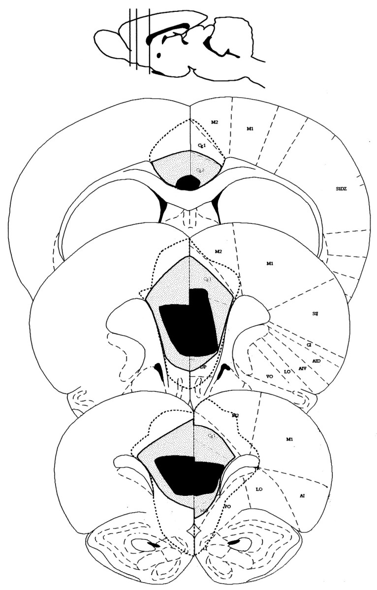
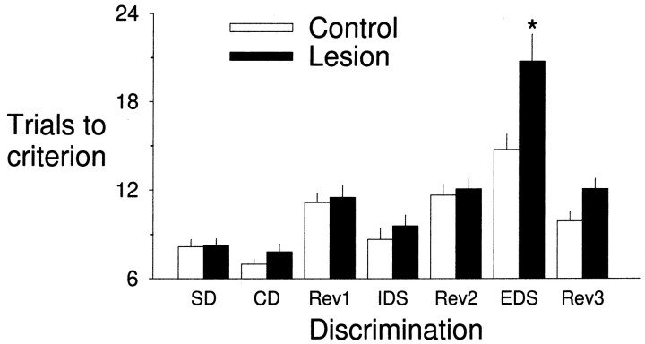
If rodents do not display the behavioral complexity that is subserved in primates by prefrontal cortex, then evolution of prefrontal cortex in the rat should be doubted. Primate prefrontal cortex has been shown to mediate shifts in attention between perceptual dimensions of complex stimuli. This study examined the possibility that medial frontal cortex of the rat is involved in the shifting of perceptual attentional set. We trained rats to perform an attentional set-shifting task that is formally the same as a task used in monkeys and humans. Rats were trained to dig in bowls for a food reward. The bowls were presented in pairs, only one of which was baited. The rat had to select the bowl in which to dig by its odor, the medium that filled the bowl, or the texture that covered its surface. In a single session, rats performed a series of discriminations, including reversals, an intradimensional shift, and an extradimensional shift. Bilateral lesions by injection of ibotenic acid in medial frontal cortex resulted in impairment in neither initial acquisition nor reversal learning. We report here the same selective impairment in shifting of attentional set in the rat as seen in primates with lesions of prefrontal cortex. We conclude that medial frontal cortex of the rat has functional similarity to primate lateral prefrontal cortex.
Figures
References
-
- Bussey TJ, Muir JL, Everitt BJ, Robbins TW. Triple dissociation of anterior cingulate, posterior cingulate, and medial frontal cortices on visual discrimination tasks using a touchscreen testing procedure for the rat. Behav Neurosci. 1997;111:920–936. - PubMed
-
- Delatour B, Gisquet-Verrier P. Lesions of the prelimbic-infralimbic cortices in rats do not disrupt response selection processes but induce delay-dependent deficits: evidence for a role in working memory? Behav Neurosci. 1999;113:941–955. - PubMed
-
- Dias R, Robbins TW, Roberts AC. Dissociation in prefrontal cortex of affective and attentional shifts. Nature. 1996a;380:69–72. - PubMed
-
- Dias R, Robbins TW, Roberts AC. Primate analogue of the Wisconsin Card Sorting Test: effects of excitotoxic lesions of the prefrontal cortex in the marmoset. Behav Neurosci. 1996b;110:872–886. - PubMed
Publication types
MeSH terms
Grants and funding
LinkOut - more resources
Full Text Sources
Other Literature Sources