. 2000 Jun 1;20(11):4345-54.
doi: 10.1523/JNEUROSCI.20-11-04345.2000.

mu-Opioid receptors often colocalize with the substance P receptor (NK1) in the trigeminal dorsal horn

Affiliations

mu-Opioid receptors often colocalize with the substance P receptor (NK1) in the trigeminal dorsal horn

S A Aicher et al. J Neurosci. .

Abstract

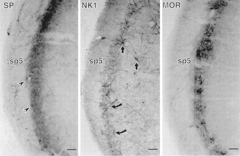
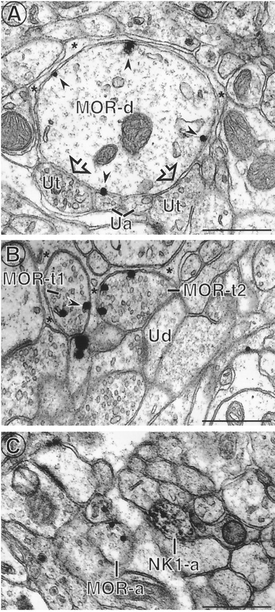
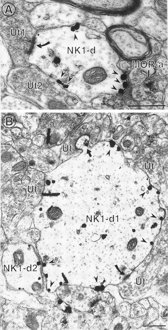
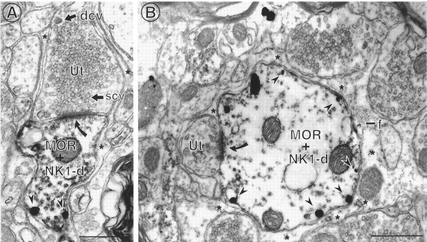
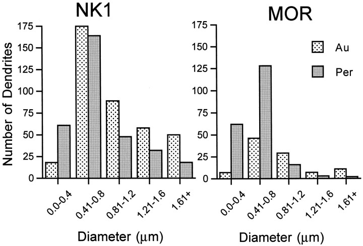
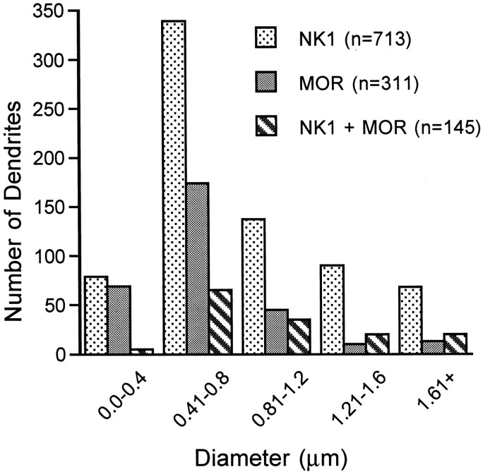
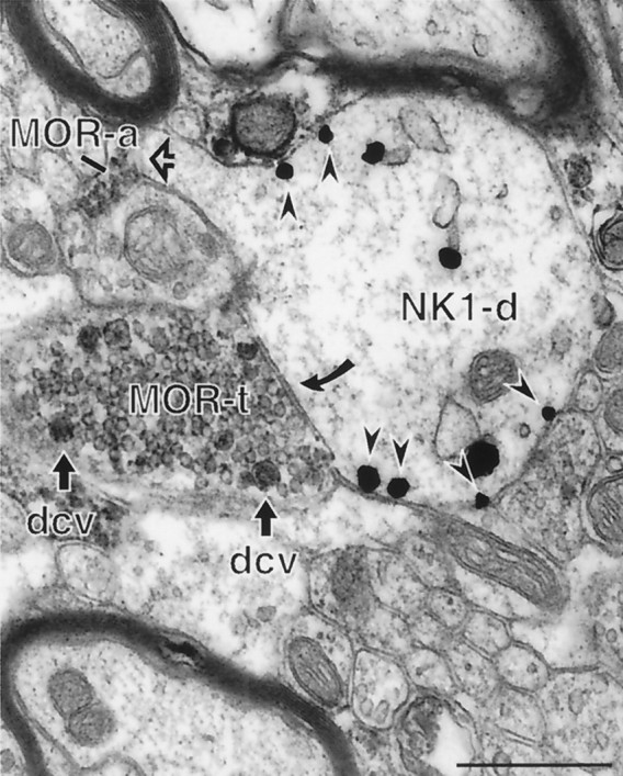
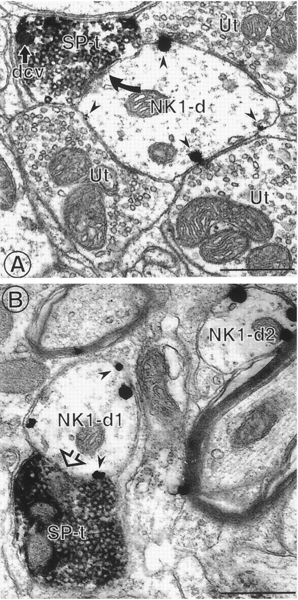
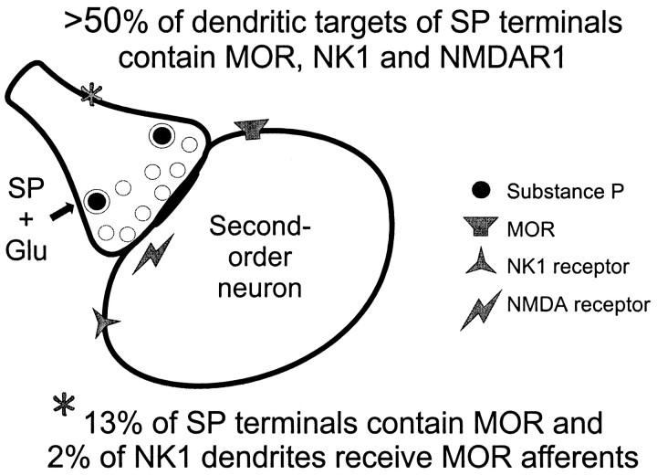
Substance P (SP) is a peptide that is present in unmyelinated primary afferents to the dorsal horn and is released in response to painful or noxious stimuli. Opiates active at the mu-opiate receptor (MOR) produce antinociception, in part, through modulation of responses to SP. MOR ligands may either inhibit the release of SP or reduce the excitatory responses of second-order neurons to SP. We examined potential functional sites for interactions between SP and MOR with dual electron microscopic immmunocytochemical localization of the SP receptor (NK1) and MOR in rat trigeminal dorsal horn. We also examined the relationship between SP-containing profiles and NK1-bearing profiles. We found that 56% of SP-immunoreactive terminals contact NK1 dendrites, whereas 34% of NK1-immunoreactive dendrites receive SP afferents. This result indicates that there is not a significant mismatch between sites of SP release and available NK1 receptors, although receptive neurons may contain receptors at sites distant from the peptide release site. With regard to opioid receptors, we found that many MOR-immunoreactive dendrites also contain NK1 (32%), whereas a smaller proportion of NK1-immunoreactive dendrites contain MOR (17%). Few NK1 dendrites (2%) were contacted by MOR-immunoreactive afferents. These results provide the first direct evidence that MORs are on the same neurons as NK1 receptors, suggesting that MOR ligands directly modulate SP-induced nociceptive responses primarily at postsynaptic sites, rather than through inhibition of SP release from primary afferents. This colocalization of NK1 and MORs has significant implications for the development of pain therapies targeted at these nociceptive neurons.


Figures

Fig. 1.
Fig. 1.
Light micrographs showing immunoperoxidase labeling for SP, NK1, and MOR in the dorsal horn of spinal trigeminal caudalis. Labeling for SP and MOR was found almost exclusively in laminae I and II of the trigeminal dorsal horn, adjacent to the spinal trigeminal tract (sp5). SP labeling was seen primarily in punctate profiles resembling axon terminals (arrowheads). NK1 labeling was seen in a few cell bodies in both lamina I and deeper laminae (straight arrows) but was primarily found in long, slender processes resembling dendrites (curved arrows). MOR immunolabeling was diffuse, and it was difficult to identify specific structures at the light microscopic level, but most of the labeled structures were seen in the laminae that contained the highest density of SP and NK1 labeling. Scale bars, 0.2 mm.
Fig. 2.
Fig. 2.
MOR immunoreactivity was detected in dendrites, axons, and axon terminals. A, A MOR-labeled dendrite (MOR-d) contains numerous immunogold particles (arrowheads) associated with the plasma membrane. A portion of the dendrite is apposed to unlabeled axon terminals (Ut) (open arrows) and unmyelinated axons (Ua), whereas the remaining surface is apposed to unlabeled astrocytic glial processes (asterisk).B, Two MOR immunogold-labeled axon terminals (MOR-t1 and MOR-t2) contain gold particles (arrowhead) associated with nonsynaptic portions of the plasma membrane. MOR-t2 is apposed to an unlabeled dendrite (Ud), but the other portions of the terminal are surrounded by unlabeled glial processes (asterisk).C, In a field containing many unmyelinated axons, an immungold-labeled unmyelinated axon (MOR-a) is near but not contacting an immunoperoxidase-labeled axon (NK1-a). Scale bars, 0.5 μm.
Fig. 3.
Fig. 3.
NK-1 immunoreactivity is primarily found in dendrites in trigeminal dorsal horn. A, An immunogold-labeled NK1 dendrite (NK1-d) contains several gold particles that are associated with nonsynaptic portions of the plasma membrane. This dendrite receives an asymmetric synapse (filled curved arrow) from an unlabeled terminal (Ut1) and is apposed to another unlabeled terminal (Ut2). A cluster of gold particles is located along the contact with Ut2, but no synaptic density is apparent between these membranes. A peroxidase-labeled MOR axon is separated from the NK1-labeled dendrite by an unlabeled axon interposed between these profiles. B, An immunogold-labeled NK1 dendrite (NK1-d) contains numerous gold particles associated with nonsynaptic portions of the plasma membrane (arrowheads). This dendrite is apposed by numerous unlabeled terminals (Ut), one of which forms an asymmetric synapse on the main part of the dendrite (filled curved arrow, bottom of figure) and one of which synapses with a small spinous extension of the dendrite (top of figure). An NK1 gold particle is located at the neck of this dendritic spine near the synapse (filled straight arrow). Another NK1-labeled dendrite (NK1-d2) contains gold particles that are exclusively associated with the plasma membrane and are all extrasynaptic in this plane of section. Scale bars, 0.5 μm.
Fig. 4.
Fig. 4.
NK1 and MOR are often contained in the same dendrite. A, A dendrite containing both MOR and NK1 (MOR + NK1-d) receives an asymmetric synapse from an unlabeled terminal that contains both small clear vesicles (scv) and dense-core vesicles (dcv). The terminal and the dendrite are surrounded by unlabeled astrocytic processes (asterisk). Immunogold particles (arrowheads) representing NK1 are located at nonsynaptic plasma membrane sites. B, A dendrite containing both immunoperoxidase labeling for MOR and immunogold labeling for NK1 receives an asymmetric synapse (filled curved arrow) from an unlabeled terminal (Ut). NK1 immunogold particles (arrowheads) are located at nonsynaptic plasma membrane sites. The remainder of the dendritic surface is surrounded by unlabeled astrocytic processes (asterisk) containing glial filaments (f). Scale bars, 0.5 μm.
Fig. 5.
Fig. 5.
Most dendrites containing NK1 or MOR alone are medium-sized. Histograms show the number of dendrites containing only NK1 (left panel) or only MOR (right panel) that were identified using either immunogold (Au) or immunoperoxidase (Per) detection methods. The cross-sectional diameter of each dendrite was classified in a 0.4 μm bin (x-axis), and the number of dendrites in each size category for each detection method is represented along the y-axis. These values are for single-labeled profiles only. Profiles <0.4 μm were less frequently detected with the immunogold method compared with immunoperoxidase for both receptor types. The majority of labeled dendrites were 0.4–0.8 μm in diameter for both receptors using either detection method. Total numbers of profiles (n): n = 390 for NK1-Au, n = 323 for NK1-Per; n= 100 for MOR-Au, n = 211 for MOR-Per.
Fig. 6.
Fig. 6.
The distribution of NK1-, MOR-, and dual-labeled profiles varies with diameter. The data are pooled for immunogold and immunoperoxidase detection methods. Dually labeled profiles <0.4 μm were rarely detected, whereas the largest number of dually labeled profiles were 0.4–0.8 μm in diameter. The number of dually labeled profiles is greater than the number of MOR single-labeled profiles at diameters >1.2 μm, whereas single-labeled NK1 profiles were more frequently detected in all size categories.
Fig. 7.
Fig. 7.
MOR-labeled axon terminals sometimes contact NK1-immunoreactive dendrites. An immunoperoxidase-labeled axon terminal (MOR-t) forms a symmetric synapse (filled curved arrow) with an immunogold-labeled dendrite (NK1-d). The MOR labeling is strongly associated with dense-core vesicles (dcv). The dendrite is also apposed to a MOR-labeled axon (MOR-a) at the head of a small spine (open arrow). Most of the immunogold particles (arrowheads) are associated with nonsynaptic portions of the plasma membrane. Scale bar, 0.5 μm.
Fig. 8.
Fig. 8.
SP-containing terminals contact NK1-immunoreactive dendrites. A, An immunoperoxidase-labeled SP terminal (SP-t) forms an asymmetric synapse (filled curved arrow) with an immunogold-labeled NK1 dendrite (NK1-d). The peroxidase labeling is located throughout the axon terminal, which contains both small clear vesicles and at least one dense-core vesicle (dcv). The immunogold particles (arrowheads) are exclusively associated with nonsynaptic portions of the plasma membrane. This NK1-labeled dendrite is also apposed to three unlabeled axon terminals (Ut) containing small clear vesicles. B, An immunogold-labeled NK1 dendrite (NK1-d1) is contacted (open arrow) by an immunoperoxidase-labeled SP terminal (SP-t). Immunogold particles (arrowheads) in this dendrite and another dendrite in the field (NK1-d2) are associated with nonsynaptic portions of the plasma membrane. Scale bars, 0.5 μm.
Fig. 9.
Fig. 9.
Schematic summary of the localization of MOR, NK1, and NMDAR1 receptors relative to SP-containing axon terminals in the dorsal horn. This model is based on results from the present study and two previous studies on the localization of SP and each receptor in this area using similar ultrastructural methods and analyses.

References

    1. Aicher SA, Reis DJ, Nicolae R, Milner TA. Monosynaptic projections from the medullary gigantocellular reticular formation to sympathetic preganglionic neurons in the thoracic spinal cord. J Comp Neurol. 1995;363:563–580. - PubMed
    1. Aicher SA, Saravay RH, Cravo SL, Jeske I, Morrison SF, Reis DJ, Milner TA. Monosynaptic projections from the nucleus tractus solitarii to C1 adrenergic neurons in the rostral ventrolateral medulla: comparison with input from the caudal ventrolateral medulla. J Comp Neurol. 1996;373:62–75. - PubMed
    1. Aicher SA, Sharma S, Cheng PY, Pickel VM. The N-methyl-d-aspartate (NMDA) receptor is postsynaptic to substance P-containing axon terminals in the rat superficial dorsal horn. Brain Res. 1997;772:71–81. - PubMed
    1. Aicher SA, Sharma S, Cheng PY, Liu-Chen LY, Pickel VM. Dual ultrastructural localization of μ-opiate receptors and substance P in the dorsal horn. Synapse. 2000;36:12–20. - PubMed
    1. Allen BJ, Rogers SD, Ghilardi JR, Menning PM, Kuskowski MA, Basbaum AI, Simone DA, Mantyh PW. Noxious cutaneous thermal stimuli induce a graded release of endogenous substance P in the spinal cord: imaging peptide action in vivo. J Neurosci. 1997;17:5921–5927. - PMC - PubMed

Publication types

MeSH terms