. 2000 Jun;130(3):527-38.
doi: 10.1038/sj.bjp.0703356.

Exploration of the ligand binding site of the human 5-HT(4) receptor by site-directed mutagenesis and molecular modeling

Affiliations

Exploration of the ligand binding site of the human 5-HT(4) receptor by site-directed mutagenesis and molecular modeling

J Mialet et al. Br J Pharmacol. 2000 Jun.

Abstract

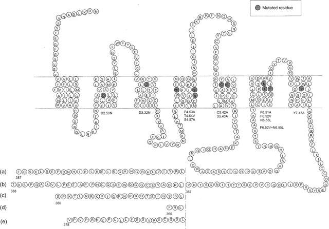
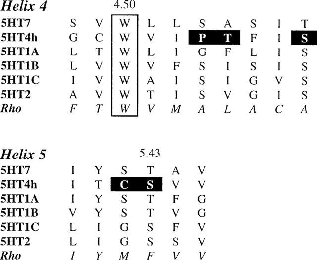
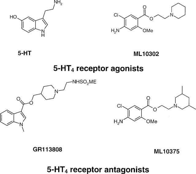
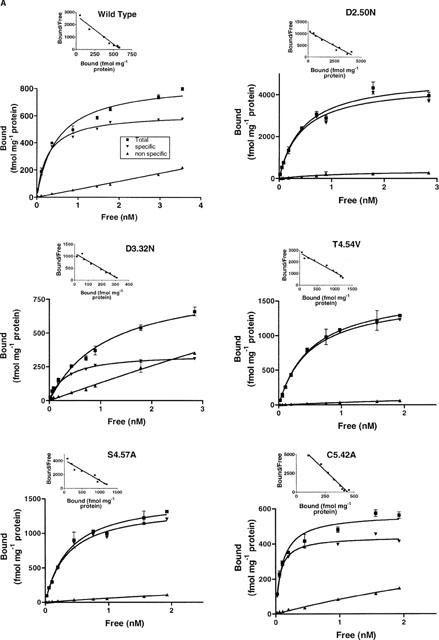
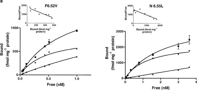
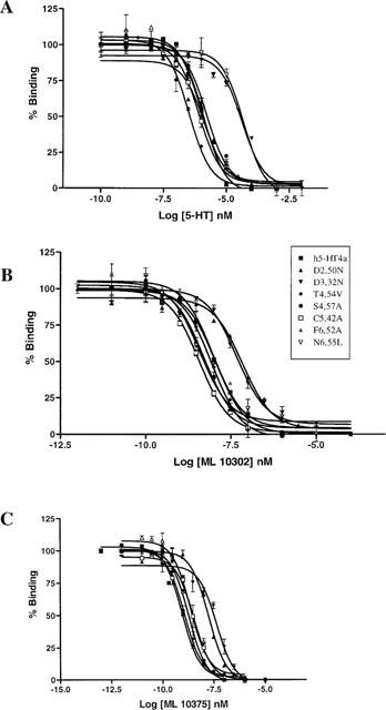
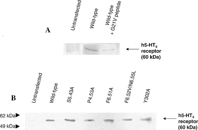
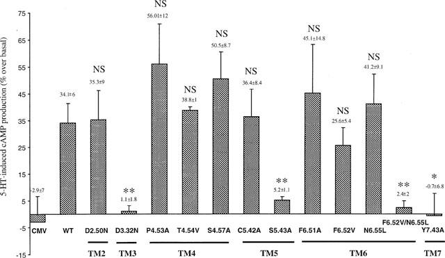
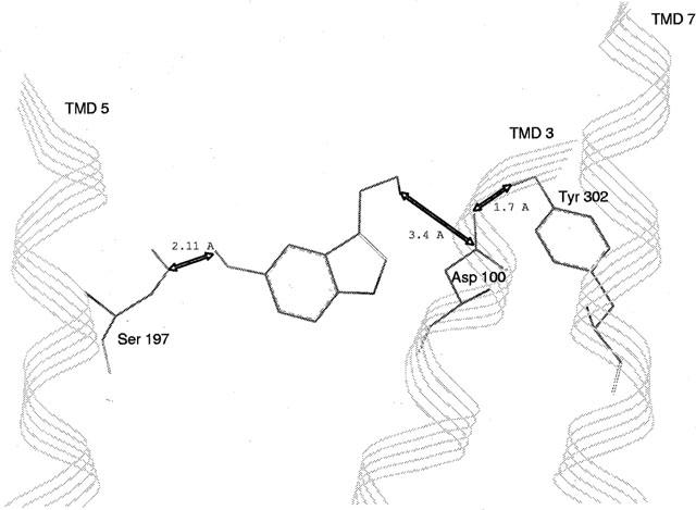
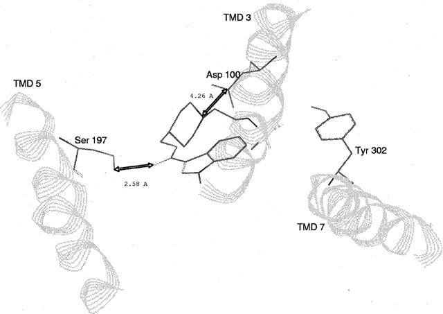
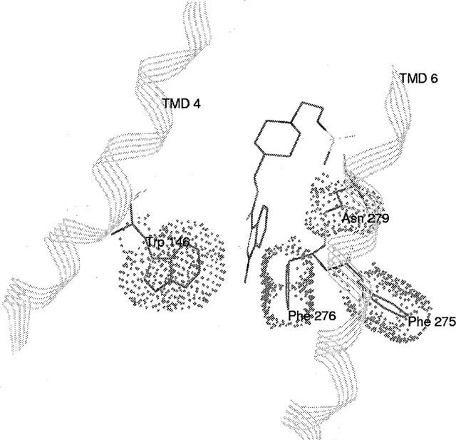
Among the five human 5-HT(4) (h5-HT(4)) receptor isoforms, the h5-HT(4(a)) receptor was studied with a particular emphasis on the molecular interactions involved in ligand binding. For this purpose, we used site-directed mutagenesis of the transmembrane domain. Twelve mutants were constructed with a special focus on the residue P4.53 of helix IV which substitutes in h5-HT(4) receptors the highly conserved S residue among the rhodopsin family receptors. The mutated receptors were transiently expressed in COS-7 cells. Ligand binding or competition studies with two h5-HT(4) receptor agonists, serotonin and ML10302 and two h5-HT(4) receptor antagonists, [(3)H]-GR113808 and ML10375 were performed on wild type and mutant receptors. Functional activity of the receptors was evaluated by measuring the ability of serotonin to stimulate adenylyl cyclase. Ligand binding experiments revealed that [(3)H]-GR113808 did not bind to mutants P4.53A, S5.43A, F6.51A, Y7.43A and to double mutant F6.52V/N6.55L. On the other hand mutations D3.32N, S5.43A and Y7.43A appeared to promote a dramatic decrease of h5-HT(4(a)) receptor functional activity. From these studies, S5.43 and Y7.43 clearly emerged as common anchoring sites to antagonist [(3)H]-GR113808 and to serotonin. According to these results, we propose ligand-receptor complex models with serotonin and [(3)H]-GR113808. For serotonin, three interaction points were selected including ionic interaction with D3.32, a stabilizing interaction of this ion pair by Y7.43 and a hydrogen bond with S5.43. [(3)H]-GR113808 was also docked, based on the same type of interactions with S5.43 and D3.32: the proposed model suggested a possible role of P4.53 in helix IV structure allowing the involvement of a close hydrophobic residue, W4.50, in a hydrophobic pocket for hydrophobic interactions with the indole ring of [(3)H]-GR113808.


Figures

Figure 1
Figure 1
Snake representation of the five isoforms of h5-HT4 receptor. The amino acid sequences diverge after Leu 358. Site-directed mutagenesis studies were performed on the h5-HT4(a) receptor isoform.
Figure 2
Figure 2
Sequence alignment of helices IV and V of 5-HT receptors. In bold are indicated the mutated residues of h5-HT4 receptor.
Figure 3
Figure 3
Chemical structures of h5-HT4 receptor agonists and antagonists selected for these studies.
Figure 4
Figure 4
Saturation analysis of [3H]-GR113808 binding to COS-7 cells transfected with wild-type or mutant human h5-HT4(a) receptors. Non-specific binding was determined with 10 μM ML10375. Experiments were performed in triplicate using a range of nine concentrations of radioligand.
Figure 4
Figure 4
Saturation analysis of [3H]-GR113808 binding to COS-7 cells transfected with wild-type or mutant human h5-HT4(a) receptors. Non-specific binding was determined with 10 μM ML10375. Experiments were performed in triplicate using a range of nine concentrations of radioligand.
Figure 5
Figure 5
Competition binding of agonists (5-HT (A), ML10302 (B)) and antagonist (ML10375 (C)) in COS-7 cells transfected with wild-type and mutant h5-HT4(a) receptors. Concentration of [3H]-GR113808 was 0.3 nM. Experiments were performed in triplicate using a range of nine concentrations of displacing ligand.
Figure 6
Figure 6
Protein immunoblotting of the mutant and wild type h5-HT4(a) receptors expressed in COS-7 cells. Plasma membrane protein extracts were separated on a polyacrylamide gel and analysed by immunoblotting with antiserum against the second extracellular loop of h5-HT4(a) receptor (see methods). (A) COS-7 cells were transfected with the wild-type h5-HT4(a) receptor (lane 2 and lane 3) or the corresponding control (lane 1). The immunoreactivity of wild-type receptor was blocked by incubation with a peptide (G21V) corresponding to the second extracellular loop of the receptor (lane 3). (B) In COS-7 cells transfected with the wild-type h5-HT4 receptor (lane 2) and different mutants (lane 3–7), a specific band migrating to the level of 60 kDa was detected whereas in non transfected control cells (lane 1) no labelling was detected at this position.
Figure 7
Figure 7
Functional response of wild-type and mutant human 5-HT4(a) receptors. Cyclic AMP accumulation in COS-7 cells expressing wild-type and mutant 5-HT4(a) receptors after stimulation with 1 μM of 5-HT. Values are expressed as percentage of basal cyclic AMP. Data are the mean±s.e.mean of 3–4 experiments performed in triplicate. CMV represents COS-7 cells transfected with crude pRC/CMV vector. WT, Wild Type; NS, Not Significant; *P<0.05, **P<0.01 with t-test between wild-type and mutant receptors.
Figure 8
Figure 8
Serotonin-h5-HT4 receptor complex model. Obtained by docking experiment, this model is characterized by distances: (i) d=2.11 Å between the hydroxylic oxygen of S197(5.43) and the serotonin hydroxylic hydrogen, (ii) d=3.4 Å between the carboxylic carbon of D100(3.32) and the serotonin basic nitrogen, and (iii) d=1.7 Å between the carboxylate oxygen in D100(3.32) and the phenolic hydrogen of Y302(4.43).
Figure 9
Figure 9
GR113808-h5-HT4 receptor complex model. It is characterized by distances: (i) d=2.58 Å between the hydroxylic hydrogen of S197(5.43) and the GR113808 carbonyl oxygen, (ii) d=4.26 Å between the carboxylic carbon of D100(3.32) and the GR113808 basic nitrogen.
Figure 10
Figure 10
Representation of hydrophobic interactions between the indol ring of GR113808 and a pocket constituted by W146(4,46), F275(6.51), F276(6.52) and N279(6.55).

References

    1. ALMAULA N., EBERSOLE B.J., BALLESTEROS J.A., WEINSTEIN H., SEALFON S.C. Contribution of a helix 5 locus selectivity of hallucinogenic and non-hallucinogenic ligands for the human 5-Hydroxytryptamine2A and 5-Hydroxytryptamine2C receptors: direct and indirect effects on ligand affinity mediated by the same locus. Mol. Pharmacol. 1996;50:34–42. - PubMed
    1. BALDWIN J.M. Structure and function of receptors coupled to G proteins. Curr. Opin. Cell. Biol. 1994;6:180–190. - PubMed
    1. BALDWIN J.M., SCHERTLER G.F.X., UNGER V.M. An alpha-carbon template for the transmembrane helices in rhodopsin family of G-protein-coupled receptors. J. Mol. Biol. 1997;272:144–164. - PubMed
    1. BALLESTEROS J.A., WEINSTEIN H. Integrated methods for modelling G-protein coupled receptors. Methods Neurosci. 1995;25:366–428.
    1. BLONDEL O., GASTINEAU M., DAHMOUNE Y., LANGLOIS M., FISCHMEISTER R. Cloning, expression, and pharmacology of four human 5-hydroxytryptamine4 receptor isoforms produced by alternative splicing in the carboxyl terminus. J. Neurochem. 1998;70:2252–2261. - PubMed

MeSH terms