. 2000 Jun;74(12):5534-41.
doi: 10.1128/jvi.74.12.5534-5541.2000.

Competing death programs in poliovirus-infected cells: commitment switch in the middle of the infectious cycle

Affiliations

Competing death programs in poliovirus-infected cells: commitment switch in the middle of the infectious cycle

V I Agol et al. J Virol. 2000 Jun.

Abstract

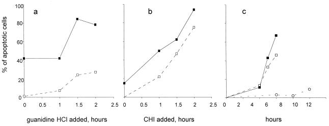
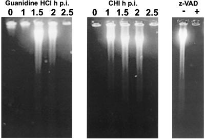
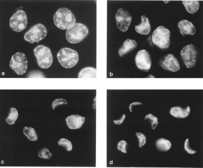
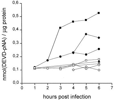
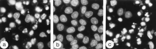
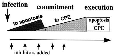
Productive poliovirus infection of HeLa cells leads to the canonical cytopathic effect (CPE), whereas certain types of abortive infection result in apoptosis. To define the time course of commitment to the different types of poliovirus-induced death, inhibitors of viral replication (guanidine HCl) or translation (cycloheximide) were added at different times postinfection (p.i.). Early in the infection (during the first approximately 2 h p.i.), predominantly proapoptotic viral function was expressed, rendering the cells committed to apoptosis, which developed several hours after viral expression was arrested. In the middle of infection, concomitantly with the onset of fast generation of viral progeny, the implementation of the viral apoptotic program was abruptly interrupted. In particular, activation of an Asp-Glu-Val-Asp (DEVD)-specific caspase(s) occurring in the apoptosis-committed cells was prevented by the ongoing productive infection. Simultaneously, the cells retaining normal or nearly normal morphology became committed to CPE, which eventually developed regardless of whether or not further viral expression was allowed to proceed. The implementation of the poliovirus-induced apoptotic program was suppressed in HeLa cells overexpressing the Bcl-2 protein, indicating that the fate of poliovirus-infected cells depends on the balance of host and viral pro- and antiapoptotic factors.


Figures

FIG. 1
FIG. 1
Development of apoptosis after interruption of productive poliovirus infection. Guanidine HCl (100 μg/ml) (a) or CHI (10 μg/ml) (b) was added to the infected HeLa cells at the indicated times, and the proportion of apoptotic cells was determined at either 12 h p.i. (solid squares) or 6 h p.i. (empty squares). In panel c, CHI (10 μg/ml) was added to the poliovirus-infected cells at 0 (circles), 1.5 (empty squares), and 2 (solid squares) h p.i., and the proportion of apoptotic cells was determined at the indicated times. Thirty minutes before fixation, Hoechst-33342 was added to the culture medium (to a final concentration of 5 μg/ml). Several low-magnification fields were photographed with a fluorescence microscope, and the proportion of the apoptotic cells was determined after counting at least 800 nuclei.
FIG. 2
FIG. 2
The switch in the cell commitment in the middle of poliovirus infection. Fluorescence microscopy of Hoechst-33342-stained HeLa cells was performed. (a) Uninfected cells treated with CHI (10 μg/ml) for 10 h; (b) infected cells at 8 h p.i. with no CHI added; and (c to f) cells to which CHI (10 μg/ml) was added 1.5 (c), 2 (d), 2.5 (e), and 3 (f) h p.i. Some of the apoptotic and CPE cells are indicated by arrowheads and arrows, respectively.
FIG. 3
FIG. 3
Electron microscopy of the cells in which poliovirus infection was interrupted at different times. Infected cells at 8 h p.i. to which CHI (10 μg/ml) was added 1.5 (a), 2 (b and c), and 3 (d) h p.i. An infected cell at 6 h p.i. of productive infection is shown in panel e. Some of the areas of condensed chromatin in both apoptotic cells (a and b) and cells exhibiting CPE (c and d) are labeled with c, and virus-induced vesicles are marked v. Bars, 1 μm.
FIG. 4
FIG. 4
DNA fragmentation in the cells with interrupted poliovirus infection. Guanidine HCl (100 μg/ml) or CHI (10 μg/ml) was added to the infected cells at the times (in hours) p.i. indicated over the lanes. CHI was added 2 h p.i. to the samples with (+) or without (−) zVAD.fmk (z-VAD) (100 μg/ml). DNA was extracted at 6 h p.i.
FIG. 5
FIG. 5
Time course of development of CPE. Productively infected cells were stained with Hoechst-33342 and fixed at 3 (a), 4 (b), 5 (c), and 8 (d) h p.i.
FIG. 6
FIG. 6
Activation of a DEVD-specific caspase(s) during apoptosis and CPE. The DEVD-specific caspase activity was determined as described in Materials and Methods in extracts from mock-infected cells (triangles), productively infected cells (circles), and infected cells to which CHI (10 μg/ml) was added 2 h (squares) and 3 h (diamonds) p.i. The enzyme activity was measured in samples without (solid symbols) and with (open symbols) zVAD.fmk (10 μg/ml) and is expressed as nanomoles of DEVD-pNA hydrolyzed per microgram of protein.
FIG. 7
FIG. 7
Suppressing effect of Bcl-2 expression on the development of poliovirus-induced apoptosis. Normal (a) and Bcl-2-expressing (b and c) HeLa cells were infected with poliovirus, and CHI (10 μg/ml) was added at 1.5 h p.i. The Hoechst-33342-stained cells were fixed at 6 (a and b) or 24 (c) h p.i., respectively.
FIG. 8
FIG. 8
Schematic representation of the time course of commitment of poliovirus-infected HeLa cells to apoptosis and CPE. The type of death and the proportion of dying cells depend on the time of addition of inhibitors of viral gene expression.

References

    1. Agol V I, Belov G A, Bienz K, Egger D, Kolesnikova M S, Raikhlin N T, Romanova L I, Smirnova E A, Tolskaya E A. Two types of death of poliovirus-infected cells: caspase involvement in the apoptosis but not cytopathic effect. Virology. 1998;252:343–353. - PubMed
    1. Bienz K, Egger D, Rasser Y, Bossart W. Kinetics and location of poliovirus macromolecular synthesis in correlation to virus-induced cytopathology. Virology. 1980;100:390–399. - PubMed
    1. Bienz K, Egger D, Rasser Y, Bossart W. Intracellular distribution of poliovirus proteins and the induction of virus-specific cytoplasmic structures. Virology. 1983;131:39–48. - PubMed
    1. Bienz K, Egger D, Troxler M, Pasamontes L. Structural organization of poliovirus RNA replication is mediated by viral proteins of the P2 genomic region. J Virol. 1990;64:1156–1163. - PMC - PubMed
    1. Bienz K, Egger D, Pfister T, Troxler M. Structural and functional characterization of the poliovirus replication complex. J Virol. 1992;66:2740–2747. - PMC - PubMed

Publication types

MeSH terms