. 2000 Jun;74(12):5577-86.
doi: 10.1128/jvi.74.12.5577-5586.2000.

Parameters of human immunodeficiency virus infection of human cervical tissue and inhibition by vaginal virucides

Affiliations
Free PMC article

Parameters of human immunodeficiency virus infection of human cervical tissue and inhibition by vaginal virucides

P Greenhead et al. J Virol. 2000 Jun.
Free PMC article

Abstract

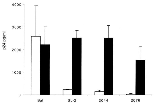
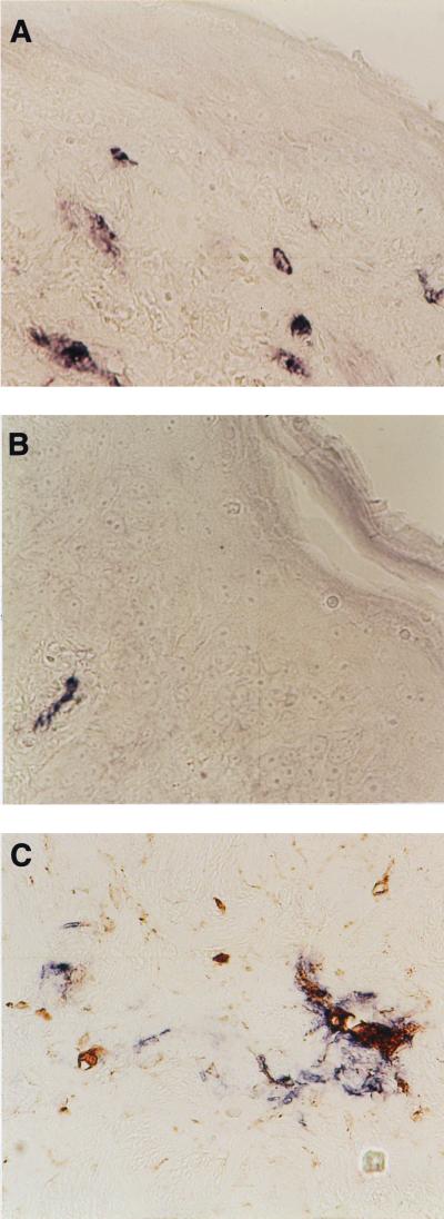
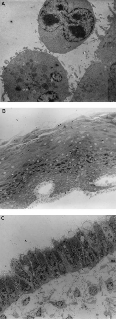
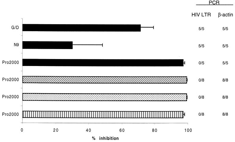
Heterosexual transmission of human immunodeficiency virus (HIV) is the most frequent mode of infection worldwide. However, the immediate events between exposure to infectious virus and establishment of infection are still poorly understood. This study investigates parameters of HIV infection of human female genital tissue in vitro using an explant culture model. In particular, we investigated the role of the epithelium and virucidal agents in protection against HIV infection. We have demonstrated that the major target cells of infection reside below the genital epithelium, and thus HIV must cross this barrier to establish infection. Immune activation enhanced HIV infection of such subepithelial cells. Furthermore, our data suggest that genital epithelial cells were not susceptible to HIV infection, appear to play no part in the transfer of infectious virus across the epithelium, and thus may provide a barrier to infection. In addition, experiments using a panel of virucidal agents demonstrated differential efficiency to block HIV infection of subepithelial cells from partial to complete inhibition. This is the first demonstration that virucidal agents designed for topical vaginal use block HIV infection of genital tissue. Such agents have major implications for world health, as they will provide women with a mechanism of personal and covert protection from HIV infection.


Figures

FIG. 1
FIG. 1
Kinetics of HIV replication in cervical explant tissue. (A) Accumulation of HIV-1 DNA in ectocervical explants was determined 7 days after infection with HIV-1BaL, HIV-1IIIB, or HIV-1RF as assayed for LTR DNA by quantitative real-time PCR as described in Materials and Methods. Explants either were prestimulated with PHA 2 days prior to HIV infection and subsequently cultured with IL-2 (filled bars) or were cultured in medium alone before and after infection (open bars). Data represents the mean and standard error of three independent experiments using paired explants from separate donors. p24 antigen release from ectocervical (B) and endocervical (C) explants was measured by ELISA. Explants were cultured either alone (open symbols, solid lines) or prestimulated with PHA (5 μg/ml) 2 days prior to viral exposure and restimulated 8 days post exposure. Stimulated explants were cultured in the presence of IL-2 (10 U/ml) (closed symbols, broken lines). Explants were exposed to HIV-1BaL (105 TCID50) without (◊) or with (⧫) (PHA, HIV-1IIIB (106 TCID50) without (□) or with (■) PHA, or HIV-1RF (106TCID50) without (○) or with (●) PHA. Data represent the mean from a minimum of three independent experiments using paired explants from separate donors. (t test; ∗, P = <0.05; ∗∗, P = <0.01).
FIG. 2
FIG. 2
Replication of primary HIV isolates in cervical explant tissue. Ectocervical explants were exposed to HIV-1 isolates SL-2 (M-tropic, NSI, CCR5 restricted), 2044 (T-tropic, SI, CXCR4 restricted), or 2076 (dualtropic, SI, able to use CCR5, CCR3, and CXCR4) by immersion in cell-free virus (103 TCID50) for 2 h at 37°C. Subsequently explant tissue was washed in PBS, and tissue was directly transferred to 96-well plates, where they were cultured in medium alone (RPMI 10%) (open bars) or stimulated by culture in the presence of PHA (5 μg/ml) for 2 days and subsequently refed with medium containing IL-2 (filled bars). Data are shown as p24 antigen release (mean ± standard error) 14 days postinfection and represent the mean from a minimum of three independent experiments using paired explants from separate donors.
FIG. 3
FIG. 3
(A and B) Immunohistochemical staining of p24 expression (blue/purple) in an ectocervical explant 7 days after infection with HIV-1BaL (A) and HIV-1RF (B). (C) Dual staining for p24 expression (blue/purple) and macrophage marker CD68 (brown) in an ectocervical explant 7 days after infection with HIV-1BaL. Original magnification for all panels, ×400.
FIG. 4
FIG. 4
(A) TEM showing lymphocyte adherence to I407 epithelial monolayer (original magnification, ×7,800). Experiments representative of a minimum of five explants for each condition. (B) TEM showing ultrastructure of stratified cervical ectocervical epithelium following exposure to PM-1 T cells infected with HIV-1BaL (original magnification, ×260). The epithelial sheet has been removed from underlying stroma following overnight treatment with dispase. Epithelial tissue was exposed to cell-associated virus for 2 h in the described diffusion chamber and gently washed before processing for TEM. There is no evidence of adherence or penetration of PM-1 T cells into the epithelium (experiment representative of three). (C) TEM showing single endocervical epithelial monolayer following exposure to PBMC infected with HIV-1BaL (original magnification, ×1,200). Epithelial cells contain multiple mucus-containing vesicles and express multiple microvilli on their apical surface. Endogenous mononuclear cells can be seen in the underlying stromal tissue. The epithelial tissue was exposed to PBMC infected with HIV-1BaL for 2 h and processed as described above. There is no evidence of adherence of paracellular migration of donor PBMC (experiment representative of five).
FIG. 5
FIG. 5
Effect of vaginal virucides on HIV-infected genital mucosa. Cervical explants were preincubated with the virucidal agent N-9 (1 μg/ml), GD (1 μg/ml), or PRO 2000 (100 μg/ml) for 1 h prior to overnight exposure to HIV-1 strain BaL (■; 105 TCID50/ml), SL2 (▨), 2044 (░⃞), or 2076 (▥) (all 103 TCID50/ml) in the continued presence of the virucidal agent. Explants were exposed to virus in a nonpolarized manner, allowing direct access to both subepithelial and epithelial cells. Following overnight exposure to viral inoculum, explants were washed extensively in PBS, stimulated by culture in the presence of PHA (5 μg/ml) for 2 days, subsequently refed with medium containing IL-2, and cultured for 14 days. Data represent percent inhibition of p24 production at day 14 of culture, compared to control cultures exposed to virus in the absence of virucidal agent, and are presented as the mean of five independent experiments using explants from separate donors. Detection of proviral DNA by PCR at the right is shown as number of experiments with detectable proviral DNA out of the total tested (limit of detection, 10 copies/explant).

References

    1. Bomsel M. Transcytosis of infectious human immunodeficiency virus across a tight human epithelial cell line barrier. Nat Med. 1997;3:42–47. - PubMed
    1. Bourinbaiar A, Lee-Huang S. Comparative in vitro study of contraceptive agents with anti-HIV activity: gramicidin, nonoxynol-9, and gossypol. Contraception. 1994;49:131–137. - PubMed
    1. Burkhard M, Obert L, O'Neil L, Diehl L, Hoover E. Mucosal transmission of cell-associated and cell-free feline immunodeficiency virus. AIDS Res Hum Retroviruses. 1997;13:347–355. - PubMed
    1. Chenine A, Matouskova E, Sanchez G, Reischig J, Pavlikova L, LeContel C, Chermann J, Hirsch I. Primary intestinal epithelial cells can be infected with laboratory-adapted strain HIV type 1 NDK but not with clinical primary isolates. AIDS Res Hum Retroviruses. 1998;14:1235–1238. - PubMed
    1. Collin M, James W, Gordon S. Development of techniques to analyse the formation of HIV provirus in primary human macrophages. Res Virol. 1991;142:105–112. - PubMed

Publication types

MeSH terms