. 2000 Jun 6;97(12):6763-8.
doi: 10.1073/pnas.100110097.

The Huntington's disease protein interacts with p53 and CREB-binding protein and represses transcription

Affiliations

The Huntington's disease protein interacts with p53 and CREB-binding protein and represses transcription

J S Steffan et al. Proc Natl Acad Sci U S A. .

Abstract

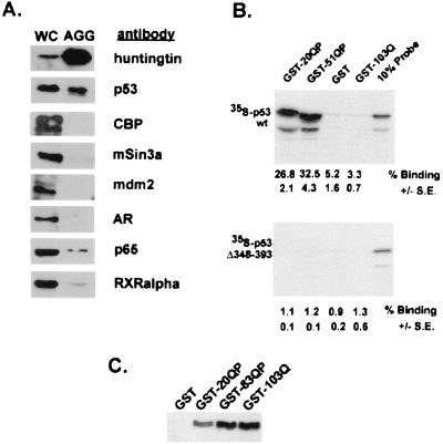
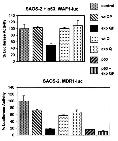
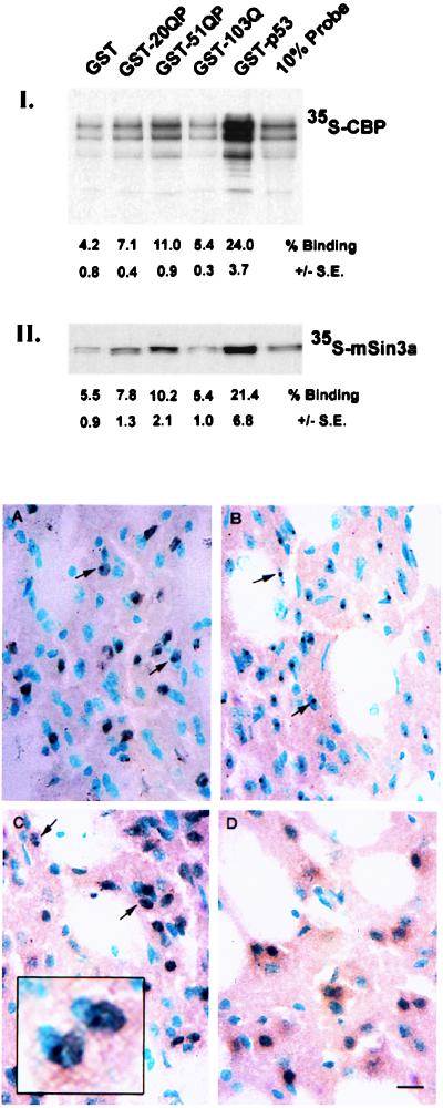
Huntington's Disease (HD) is caused by an expansion of a polyglutamine tract within the huntingtin (htt) protein. Pathogenesis in HD appears to include the cytoplasmic cleavage of htt and release of an amino-terminal fragment capable of nuclear localization. We have investigated potential consequences to nuclear function of a pathogenic amino-terminal region of htt (httex1p) including aggregation, protein-protein interactions, and transcription. httex1p was found to coaggregate with p53 in inclusions generated in cell culture and to interact with p53 in vitro and in cell culture. Expanded httex1p represses transcription of the p53-regulated promoters, p21(WAF1/CIP1) and MDR-1. httex1p was also found to interact in vitro with CREB-binding protein (CBP) and mSin3a, and CBP to localize to neuronal intranuclear inclusions in a transgenic mouse model of HD. These results raise the possibility that expanded repeat htt causes aberrant transcriptional regulation through its interaction with cellular transcription factors which may result in neuronal dysfunction and cell death in HD.


Figures

Figure 1
Figure 1
p53 colocalizes with expanded httex1p in inclusions in mammalian cell culture and interacts with httex1p in vitro and in cell culture. (A) Western blot of solubilized proteins purified from aggregates generated in HEK293 cells expressing 103Q-GFP. Results were identical to aggregates purified from 103QP-GFP (data not shown). WC denotes the whole-cell fraction from transfected cells and AGG the aggregate preparation. Equivalent ratios of WC:AGG were run on 8% SDS gels and Western blotted. Antisera were used to examine relative levels of 103Q-GFP htt, p53, CBP, mSin3a, mdm2, AR, NF-κB p65, and RXRα in the aggregate preparation. No immunoreactive htt protein was present in samples corresponding to the aggregate fraction isolated from 293 cells expressing 25Q-GFP (data not shown). (B) Binding of [35S]methionine-labeled p53(1–393) and p53(1–347) to GST-htt fusion proteins. Equivalent amounts of [35S]methionine-labeled p53(1–393) and p53(1–347) were mixed with GST-20QP, GST-51QP, GST, and GST-103Q attached to glutathione-agarose beads, incubated, and washed. The labeled proteins bound to the beads were analyzed by SDS-PAGE. Each experiment was done in triplicate. Percent binding ± SE was calculated by phosophorimager analysis and is listed below each lane. Ten percent of the input of the labeled proteins was also analyzed as shown. Slight alteration in the pattern of p53 migration in the GST-51QP lane as compared with GST-20QP lane is because of large amounts GST-51QP comigrating with labeled p53. (C) Coprecipitation of p53 with httex1p in HEK293 cells. HEK293 cells were transiently transfected with plasmids encoding p53 and either GST, GST-20QP, GST-83QP, or GST-103Q. Cells were broken and lysates were incubated with glutathione-agarose beads. After extensive washing, beads were loaded directly on an 8% SDS gel. Western blot analysis using monoclonal anti-p53 antibody is shown.
Figure 2
Figure 2
Expanded httex1p represses transcription. (Upper) SAOS-2 cells were transfected with vector alone (control) or plasmids encoding 25QP-GFP, 103QP-GFP, 25Q-myc, or 103Q-myc proteins and plasmids encoding WAF1-luciferase and p53. (Lower) SAOS-2 cells were transfected with vector alone (control) or plasmids encoding 20QP, 93QP, 25Q-myc, or 103-myc proteins and the MDR1-luciferase reporter plasmid. A p53-encoding plasmid was also transfected as indicated. Each experiment was done in triplicate. Luciferase units per milligram of protein were calculated and are demonstrated graphically as percent of vector control values.
Figure 3
Figure 3
CBP and mSin3a bind to httex1p in vitro and CBP localizes to nuclear inclusions in R6/2 mouse brains. (Top) Binding of [35S]methionine-labeled CBP or mSin3a to GST-htt fusion proteins. Equivalent amounts of [35S]methionine-labeled CBP (I) or mSin3a (II) were mixed with GST, GST-20QP, GST-51QP, GST-103Q, or GST-p53 attached to glutathione-agarose beads, washed, and the labeled proteins bound to the beads were analyzed by SDS-PAGE. Each experiment was done in triplicate. Percent binding ± SE was calculated by phosphorimager analysis and are listed below each lane. Ten percent of the input of the labeled proteins was also analyzed as shown. (A–D) Localization of CBP to neuronal intranuclear inclusions in R6/2 mice. Striatal sections from R6/2 (A–C) and littermate controls (D) were immunolabeled with anti-htt (A), -ubiquitin (B), and -CBP (C and D) antibodies. Nuclei were counterstained with methyl green. Nuclear inclusions are indicated by arrows. Scale bar, 15 μm.

Comment in

References

    1. Harper P S, editor. Huntington's Disease. London: W. B. Saunders; 1991. pp. 141–178.
    1. The Huntington's Disease Collaborative Research Group. Cell. 1993;72:971–983. - PubMed
    1. DiFiglia M, Sapp E, Chase K, Schwarz C, Meloni A, Yound C, Martin E, Vonsattel J-P, Carraway R, Reeves S A, Boyce F M, Aronin N. Neuron. 1995;14:1075–1081. - PubMed
    1. Gusella J F, MacDonald M E. Semin Cell Biol. 1995;6:21–28. - PubMed
    1. Mangiarini L, Sathasivam K, Seller M, Cozens B, Harper A, Hetherington C, Lawton M, Trottier Y, Lehrach H, Davies S W, Bates G P. Cell. 1996;87:493–506. - PubMed

Publication types