. 2000 Jun 6;97(12):6481-6.
doi: 10.1073/pnas.100340697.

CpG methylation as a mechanism for the regulation of E2F activity

Affiliations

CpG methylation as a mechanism for the regulation of E2F activity

M R Campanero et al. Proc Natl Acad Sci U S A. .

Abstract

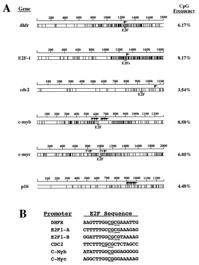
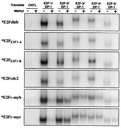
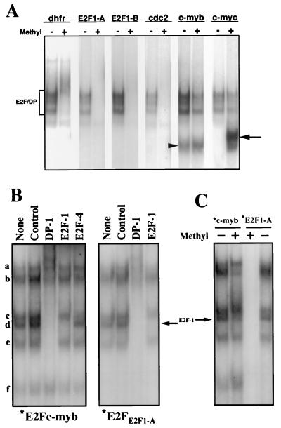
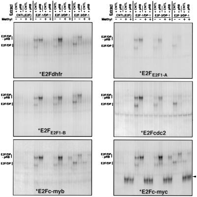
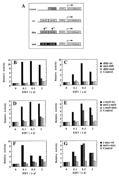
Regulation of gene expression in mammals through methylation of cytosine residues at CpG dinucleotides is involved in the development and progression of tumors. Because many genes that are involved in the control of cell proliferation are regulated by members of the E2F family of transcription factors and because some E2F DNA-binding sites are methylated in vivo, we have investigated whether CpG methylation can regulate E2F functions. We show here that methylation of E2F elements derived from the dihydrofolate reductase, E2F1, and cdc2 promoters prevents the binding of all E2F family members tested (E2F1 through E2F5). In contrast, methylation of the E2F elements derived from the c-myc and c-myb promoters minimally affects the binding of E2F2, E2F3, E2F4, and E2F5 but significantly inhibits the binding of E2F1. Consistent with these studies, E2F3, but not E2F1, activates transcription through methylated E2F sites derived from the c-myb and c-myc genes whereas both E2F1 and E2F3 fail to transactivate a reporter gene that is under the control of a methylated dihydrofolate reductase E2F site. Together, these data illustrate a means through which E2F activity can be controlled.


Figures

Figure 1
Figure 1
(A) CpG density maps of the E2F-regulated promoters dhfr, E2F1, cdc2, c-myb, and c-myc. Map plots are of the dhfr, E2F1, cdc2, c-myb, c-myc, and p16 (included as a reference) 5′ gene sequences, indicating the position of each CpG dinucleotide (vertical lines) and its frequency. The position of the E2F elements and transcription initiation sites (flags) are indicated. The two E2F elements within the E2F1 promoter are only 5 bp apart and are represented by only one bracket. P1 and P2 denote the two known c-myc promoters. The size of the analyzed gene sequences is indicated in base pairs above each map. (B) Alignment of E2F elements from the dhfr, E2F1, cdc2, c-myb, and c-myc promoters. The CpG dinucleotides within each element are underlined.
Figure 2
Figure 2
Binding of in vitro translated DP1/E2Fs to different E2F elements is regulated by CpG methylation. DP1 and either E2F1, E2F2, E2F3, E2F4, or E2F5 were co-translated in vitro, and the interaction of the resulting heterodimers with the E2Fdhfr, E2FE2F1-A, E2FE2F1-B, E2Fcdc2, E2Fc-myb, and E2Fc-myc radiolabeled probes was analyzed by EMSA. Before labeling, the DNA probes were either methylated with Sss I (+) or control methylated (−). No cDNA was added to the control (CNTL) in vitro transcription-translation reaction. Only the retarded E2F/DNA complexes are shown.
Figure 3
Figure 3
Binding of endogenous E2F factors to different E2F elements is regulated by CpG methylation. (A) Nuclear extracts from the B-lymphoblastoid cell line, X50-7, were incubated with the indicated methylated (+), or control methylated (−), radiolabeled E2F elements in the presence of sodium deoxycholate (to disrupt E2F/pRb-family member interactions). E2F-DNA complex formation was analyzed by EMSA. The position of free E2F/DP heterodimers is indicated by a bracket. The arrowhead and the arrow indicate specific complexes that bind only the E2Fc-myb probe and the methylated E2Fc-myc probe, respectively. (B) Nuclear extracts from S-phase synchronized NIH 3T3 cells were incubated with the indicated radiolabeled E2F elements. Extracts were preincubated in the absence (None) or in the presence of the indicated antibodies for 20 min before the addition of the labeled probes. Control, normal rabbit serum. (C) Nuclear extracts from S-phase synchronized NIH 3T3 cells were incubated with the indicated methylated (+) or control methylated (−) radiolabeled probes. The arrow indicates the position of the free DP1/E2F1 heterodimer.
Figure 4
Figure 4
Association of the retinoblastoma protein with E2F family members does not affect the specificity of binding to methylated E2F elements. U2OS cells were transfected with either E2F1/DP1, E2F2/DP1, E2F3/DP1, or the corresponding empty vector [CNTL(E2F)], together with either a retinoblastoma expression vector (+pRB) or the corresponding control vector (CNTL). Nuclear extracts from transfected cells were analyzed by EMSA employing the indicated methylated (+), or control methylated (−), radiolabeled E2F probes (*). The arrowhead points to a complex that binds uniquely to the methylated E2Fc-myc probe. Because unmethylated E2Fc-myc also binds a non-E2F factor that co-migrates with a free E2F form (M.R.C. and E.K.F., unpublished observations), the experiment shown in the E2Fc-myc panel was performed in the presence of a 100 molar excess of a cold competitor oligonucleotide that specifically binds this factor but not E2F proteins.
Figure 5
Figure 5
E2F3, but not E2F1, efficiently activates transcription through a subset of methylated E2F-DNA binding sites. (A) Schematic representation of the reporter plasmids used. An irrelevant DNA sequence (Control) or two copies of wild-type and methylated E2F elements from the dhfr (dhfr-wt and met-dhfr), c-myb (c-myb-wt and met-c-myb), and c-myc (c-myc-wt and met-c-myc) genes were ligated immediately upstream to the β-globin TATA box. Mutant E2F binding sites from the dhfr (dhfr-mut) and c-myb (c-myb-mut) genes that do not interact with E2F proteins were used as additional controls. Note that, although the c-myb mutant E2F element does not bind E2F factors, it retains the ability to bind another factor that interacts with a region overlapping the E2F-binding site (Fig. 3; ref. 24). Because the unmethylated and methylated wild-type c-myb E2F site similarly bind this factor, this c-myb-mut is, therefore, a proper control. E2F-BS, E2F binding site. (BG) Saos-2 cells were transfected with the indicated amounts of DP1/E2F1 (E2F1) and DP1/E2F3 (E2F3) expression vectors together with 0.5 μg of pRc-CMV-β-gal and ≈1 μg of the indicated luciferase reporter vectors described in A. Luciferase values from a representative experiment (normalized for β-galactosidase activity) are represented for each luciferase vector relative to cells transfected in the absence of E2F/DP expression vectors.

References

    1. Bird A P. Nature (London) 1986;321:209–213. - PubMed
    1. Cross S H, Bird A P. Curr Opin Genet Dev. 1995;5:309–314. - PubMed
    1. Cho K R, Hedrick L. Curr Top Microbiol Immunol. 1997;221:149–176. - PubMed
    1. Jones P A. Cancer Res. 1996;56:2463–2467. - PubMed
    1. Laird P W, Jaenisch R. Hum Mol Genet. 1994;3:1487–1495. - PubMed

Publication types

MeSH terms

LinkOut - more resources