. 2000 Jun 6;97(12):6850-5.
doi: 10.1073/pnas.110035297.

Amphetamine-induced loss of human dopamine transporter activity: an internalization-dependent and cocaine-sensitive mechanism

Affiliations

Amphetamine-induced loss of human dopamine transporter activity: an internalization-dependent and cocaine-sensitive mechanism

C Saunders et al. Proc Natl Acad Sci U S A. .

Abstract

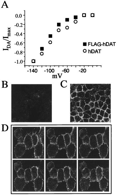
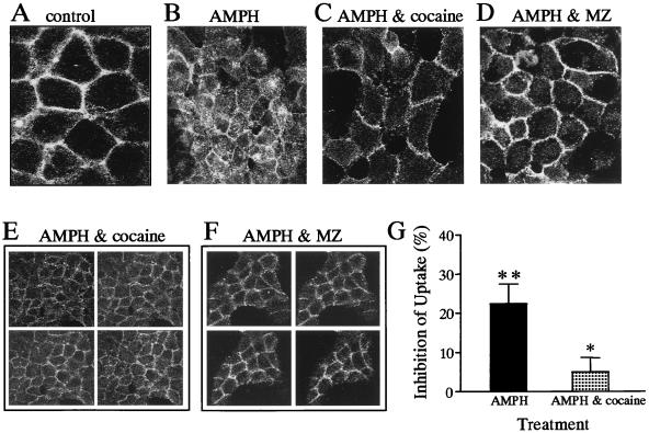
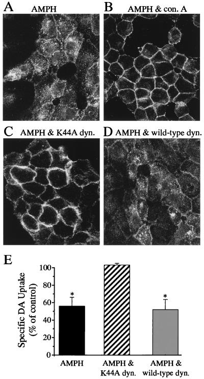
The dopamine transporter (DAT) is a target of amphetamine (AMPH) and cocaine. These psychostimulants attenuate DAT clearance efficiency, thereby increasing synaptic dopamine (DA) levels. Re-uptake rate is determined by the number of functional transporters at the cell surface as well as by their turnover rate. Here, we present evidence that DAT substrates, including AMPH and DA, cause internalization of human DAT, thereby reducing transport capacity. Acute treatment with AMPH reduced the maximal rate of [(3)H]DA uptake, decreased AMPH-induced currents, and significantly redistributed the immunofluorescence of an epitope-tagged DAT from the plasma membrane to the cytosol in human embryonic kidney 293 cells. Conversely, DAT inhibitors, such as cocaine, mazindol, and nomifensine, when administered with AMPH, blocked the reduction in [(3)H]DA uptake and the redistribution of DAT immunofluorescence to the cytosol. The reductions of [(3)H]DA uptake and AMPH-induced DAT internalization also were inhibited by coexpression of a dominant negative mutant of dynamin I (K44A), indicating that endocytosis modulates transport capacity, likely through a clathrin-mediated pathway. With this mechanism of regulation, acute application of AMPH would reduce DA uptake not only by direct competition for uptake, but also by reducing the available cell-surface DAT. Moreover, AMPH-induced internalization might diminish the amount of DAT available for DA efflux, thereby modulating the cytotoxic effects of elevated extracellular DA.


Figures

Figure 1
Figure 1
Transporter function and expression of the FLAG-hDAT cells. (A) Current-voltage relationships of the DA-induced whole-cell currents from either hDAT (○) or FLAG-hDAT (■) cells. The DA-induced current is defined as the whole-cell steady-state current recorded upon DA bath application minus the current obtained in the presence of DA and MZ at a defined membrane voltage. The membrane potential was held at −40 mV, stepping the voltage between −140 and 0 mV for 500 msec, with an interval of 4 sec. Data were normalized at −140 mV. Confocal microscopy images of hDAT cells reveal no significant fluorescence (B), whereas confocal images of the FLAG-hDAT cells show a strong cell surface staining pattern (C). Z sections of the FLAG-hDAT (D), 1 μm in thickness, go from the top to the bottom of the cell; no intracellular immunofluorescence was seen in any of the six focal planes.
Figure 2
Figure 2
AMPH-induced loss of hDAT function and hDAT cell surface expression. (A) Whole-cell currents recorded from FLAG-hDAT cells. In this representative experiment, the cell was held at −40 mV and then the voltage was stepped to −120 mV for 500 msec. The labels refer to: control current before addition of substrate (control), current after addition of 2 μM AMPH to the bath (AMPH), and inhibited current after addition of 5 μM MZ to the bath (AMPH & MZ) with AMPH still present. (B) AMPH induced a loss of hDAT whole-cell current over time. Cells were held at −40 mV and then the voltage was stepped to −120 mV for 500 msec every 1–2 min. Upon AMPH bath application, the hDAT inward current increased, reaching the maximum value over a time of 1–2 min. After several minutes of stability, the AMPH-induced current began to decrease. From this point, the AMPH-induced current recorded at −120 mV was plotted against time (each symbol type represents a single cell; n = 4). The current recorded during the different experimental time points was normalized to the AMPH-induced current recorded at a virtual time 0 (2 min before the onset of the decreasing phase). (C) Confocal microscopy images in the absence (control) and presence of 2 μM AMPH added for 1 h. The galleries are of 1-μm sections of patches of cells or single cells treated with AMPH showing extensive intracellular immunofluorescence compared with control conditions.
Figure 3
Figure 3
AMPH-induced loss of cell surface FLAG-hDAT was inhibited by blockers of hDAT activity. Confocal microscopy images of FLAG-hDAT in the presence of vehicle (A), 2 μM AMPH (B), 2 μM AMPH and 3 μM cocaine (C and E), and 2 μM AMPH and 3 μM MZ (D and F). The galleries (E and F) are 1-μm z-sections of patches of FLAG-hDAT from the top to the bottom of the cells. (G) Uptake studies of [3H]DA were done in the absence and presence of 2 μM AMPH and in the presence of 2 μM AMPH and 3 μM cocaine. AMPH significantly inhibited [3H]DA uptake, and this effect was significantly attenuated by coincubation with cocaine (* = P < 0.05; ** = P < 0.01 by paired Student's t test). [3H]DA uptake was expressed as a percentage of inhibition of uptake under control conditions. The data are the mean ± SEM of five experiments performed in triplicate.
Figure 4
Figure 4
AMPH-induced internalization of FLAG-hDAT is dynamin dependent. Confocal microscopy images of FLAG-hDAT cells incubated in the presence of either 2 μM AMPH for 1 h (A) or 2 μM AMPH plus 250 μg/ml Con A (B). Confocal microscopy images from FLAG-hDAT cells transiently transfected with either the dominant negative dynamin I mutant, K44A (C), or the wild-type dynamin (D) in the presence of 2 μM AMPH. (E) Uptake of [3H]DA (at 10°C), expressed as a percent of control, after 1 h preincubation with 2 μM AMPH in FLAG-hDAT cells, FLAG-hDAT cells transiently expressing the mutant dynamin, K44A, or the wild-type dynamin. The data are the mean ± SEM of three different experiments performed in triplicate (* = P < 0.05 by paired Student's t test).

References

    1. Iversen L L. Br J Pharmacol. 1971;41:571–591. - PMC - PubMed
    1. Giros B, Caron M G. Trends Pharmacol Sci. 1993;14:43–49. - PubMed
    1. Giros B, Jaber M, Jones S R, Wightman R M, Caron M G. Nature (London) 1996;379:606–612. - PubMed
    1. Koob G F, Bloom F E. Science. 1988;242:715–723. - PubMed
    1. Harris J E, Baldessarini R J. Neuropharmacology. 1973;12:669–679. - PubMed

Publication types

LinkOut - more resources