. 2000 Jun 6;97(12):6710-5.
doi: 10.1073/pnas.110463197.

Shear stress-induced apoptosis of adherent neutrophils: a mechanism for persistence of cardiovascular device infections

Affiliations

Shear stress-induced apoptosis of adherent neutrophils: a mechanism for persistence of cardiovascular device infections

M S Shive et al. Proc Natl Acad Sci U S A. .

Abstract

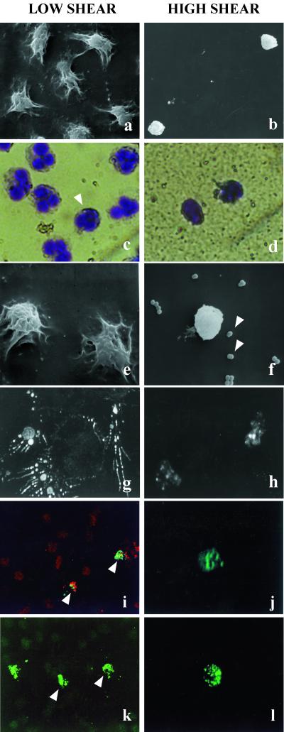
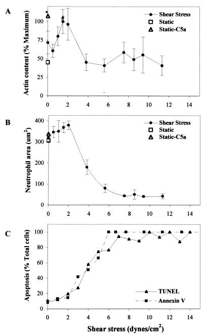
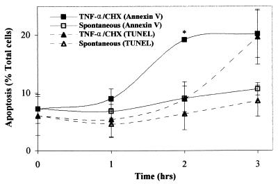
The mechanisms underlying problematic cardiovascular device-associated infections are not understood. Because the outcome of the acute response to infection is largely dependent on the function of neutrophils, the persistence of these infections suggests that neutrophil function may be compromised because of cellular responses to shear stress. A rotating disk system was used to generate physiologically relevant shear stress levels (0-18 dynes/cm(2); 1 dyne = 10 microN) at the surface of a polyetherurethane urea film. We demonstrate that shear stress diminishes phagocytic ability in neutrophils adherent to a cardiovascular device material, and causes morphological and biochemical alterations that are consistent with those described for apoptosis. Complete neutrophil apoptosis occurred at shear stress levels above 6 dynes/cm(2) after only 1 h. Morphologically, these cells displayed irreversible cytoplasmic and nuclear condensation while maintaining intact membranes. Analysis of neutrophil area and filamentous actin content demonstrated concomitant decreases in both cell area and actin content with increasing levels of shear stress. Neutrophil phagocytosis of adherent bacteria decreased with increasing shear stress. Biochemical alterations included membrane phosphatidylserine exposure and DNA fragmentation, as evaluated by in situ annexin V and terminal deoxynucleotidyltransferase-mediated dUTP end labeling (TUNEL) assays, respectively. The potency of the shear-stress effect was emphasized by comparative inductive studies with adherent neutrophils under static conditions. The combination of tumor necrosis factor-alpha and cycloheximide was ineffective in inducing >21% apoptosis after 3 h. These findings suggest a mechanism through which shear stress plays an important role in the development of bacterial infections at the sites of cardiovascular device implantation.


Figures

Figure 1
Figure 1
Effects of 60-min exposure to low (0–2 dynes/cm2) and high (>6 dynes/cm2) shear stress on neutrophils adherent to polyetherurethane urea. (a and b) Shear stress causes major morphological changes in neutrophils. Representative scanning electron micrographs demonstrate that adherent neutrophils exposed to low shear were primarily well spread, in contrast to those under high shear levels, which showed condensed and irregular morphologies. (×1,000.) (c and d) May-Grünwald/Giemsa staining revealed that neutrophils under low shear exhibited characteristic multilobed nuclei, with an occasional condensed nucleus (arrowhead in c). Nuclear condensation was observed in all cells exposed to high shear levels. (×100.) (e and f) Shear stress affects the phagocytic ability of adherent neutrophils. Representative scanning electron micrographs show that neutrophils exposed to low shear effectively located and phagocytosed preseeded S. epidermidis. Neutrophils under high shear apparently were incapable of interacting with the bacteria. Arrowheads in f show numerous bacteria in close proximity to a condensed neutrophil under high shear. (g and h) Neutrophil F-actin distribution is influenced by shear stress. F-actin was labeled with rhodamine phalloidin and visualized by using confocal microscopy. (×3,300.) Spread neutrophils in areas of low shear localized actin in pseudopodia, whereas neutrophils exposed to high shear exhibited compact actin distribution and low-intensity staining. (g, ×60, and h, ×3, computerized zoom.) (i and j) Membrane PS is exposed on neutrophils under shear stress. Syto17-counterstained (red) adherent neutrophils exposing membrane PS were identified with annexin V-FITC (green). Areas of low shear contained few annexin V-positive neutrophils, whereas high-shear areas contained sparse but exclusively annexin V-positive neutrophils. (i, ×60, and j, ×3, computerized zoom.) (k and l) DNA fragmentation also was identified after shear stress exposure. By using TUNEL-fluorescein labeling, low numbers of neutrophils with fragmentation were observed under low shear, whereas a high percentage of the sparse cells under high shear exhibited fragmentation (k, ×60, and l, ×3, computerized zoom.)
Figure 2
Figure 2
Effects of 60-min exposure to shear stress on neutrophils adherent to polyetherurethane urea. (A) F-actin content is modulated by shear stress. The maximum actin content detected through confocal analysis at 1.5 dynes/cm2 was significantly greater than were actin levels at 3.8, 5.6, and 11.3 dynes/cm2. ∗, P < 0.04. Additionally, static controls incubated with C5a demonstrated significant increases in F-actin content. ∗, P < 0.05 as compared with static controls without C5a or 3.8 and 5.6 dynes/cm2 shear levels. (B) Neutrophil area decreased with increasing shear stress. When compared with areas at all shear levels of 3.8 dynes/cm2 or greater, direct measurements produced significant differences in neutrophil area for static controls with and without C5a (P < 0.05) and shear levels of 0, 1, and 2 dynes/cm2 (P < 0.02). (C) Shear stress produced a rapid and synchronous progression from PS exposure to DNA fragmentation, as determined by in situ annexin V and TUNEL assays, respectively. Annexin V binding increased significantly for all shear levels at and above 3 dynes/cm2 as compared with 0, 1, and 2 dynes/cm2 (P < 0.05). Similarly, TUNEL-positive apoptosis was significantly greater at all shear levels of 4 dynes/cm2 or above when compared with 0, 1, 2, and 3 dynes/cm2 (P < 0.007). Data represent means ± SEM in A and B. Multiple experiments were completed separately for actin/area (9), annexin V (5), and TUNEL (7) studies, using neutrophils from at least three different blood donors.
Figure 3
Figure 3
Chemical induction of apoptosis in neutrophils adherent to static polyetherurethane urea. The combination of TNF-α and CHX induced asynchronous exposure of PS and DNA fragmentation, but was unable to produce apoptotic proportions over 20.2% even after 3 h. ∗, P < 0.04 as compared with spontaneous apoptosis at 1, 2, and 3 h. Data represent means ± SEM. Experiments were completed in duplicate with three different blood donors.

References

    1. Bandyk D, Esses G E. Surg Clin N Am. 1994;74:571–590. - PubMed
    1. Black M M. J Med Engl Tech. 1995;19:151–157. - PubMed
    1. Sugarman B, Young E J. Infect Dis Clin N Am. 1989;3:187–197. - PubMed
    1. Zimmerli W, Lew P, Waldvogel F. J Clin Invest. 1984;73:1191–2000. - PMC - PubMed
    1. Christensen G D, Simpson W A, Bisno A L, Beachey E H. Infect Immun. 1983;40:407–410. - PMC - PubMed

Publication types

LinkOut - more resources