. 2000 Jun 6;97(12):6527-31.
doi: 10.1073/pnas.120048697.

Atomic force microscopy reveals the mechanical design of a modular protein

Affiliations

Atomic force microscopy reveals the mechanical design of a modular protein

H Li et al. Proc Natl Acad Sci U S A. .

Abstract

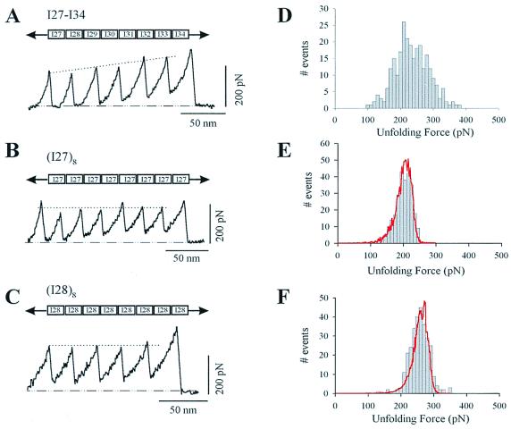
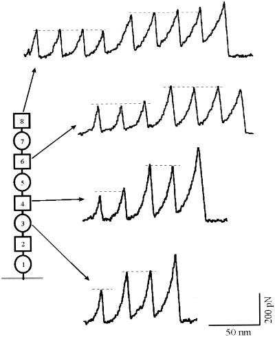
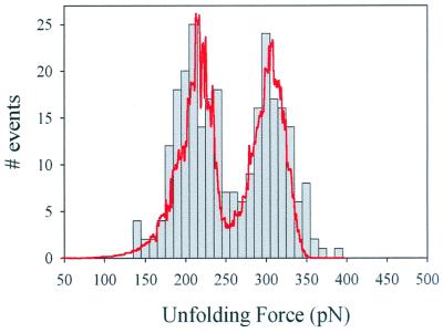
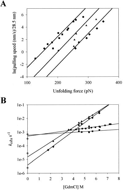
Tandem modular proteins underlie the elasticity of natural adhesives, cell adhesion proteins, and muscle proteins. The fundamental unit of elastic proteins is their individually folded modules. Here, we use protein engineering to construct multimodular proteins composed of Ig modules of different mechanical strength. We examine the mechanical properties of the resulting tandem modular proteins by using single protein atomic force microscopy. We show that by combining modules of known mechanical strength, we can generate proteins with novel elastic properties. Our experiments reveal the simple mechanical design of modular proteins and open the way for the engineering of elastic proteins with defined mechanical properties, which can be used in tissue and fiber engineering.


Figures

Figure 1
Figure 1
Force-extension curves for various recombinant fragments of human cardiac I band titin, measured with single-protein AFM techniques. (A) Force-extension curve for a recombinant protein composed of the I27-I34 region of I band titin. Notice the steady rise in the force peaks, indicating the hierarchical unfolding of its modules. By contrast, stretching either an I27 polyprotein (B) or an I28 polyprotein (C) produces sawtooth patterns with a relatively constant unfolding force. (D) Histogram of unfolding forces for the I27-I34 protein shows a broad peak spanning a ≈300 pN range of unfolding forces (E, F). Unfolding force frequency histograms for I27 and I28 polyproteins show a single peak at 204 ± 26 pN (n = 266) and 257 ± 27 pN (n = 245), respectively. The lines correspond to Monte Carlo simulations of the mean unfolding forces (10,000 trials) of eight domains placed in series by using a pulling rate of 0.6 nm/ms and an unfolding distance, Δxu, of 0.25 nm for both domains. The unfolding rate constants were ku0, 3.3 × 10⁻⁴ s−1 and 2.8 × 10⁻⁵ s−1 for I27 and I28, respectively.
Figure 2
Figure 2
Force-extension curves for a heteropolyprotein constructed as (I27-I28)4. The force-extension curves always show two distinct levels of unfolding forces (dashed lines). However, the number of modules unfolding in each case varies because the AFM tip picks up the proteins at a random location resulting in the stretch unfolding of different number of modules every time a new molecule is picked up. The diagram on the left provides an explanation for the recordings and marks the number of modules that will unfold in each case (circles represent I28 modules, squares represent I27 modules). Some exclusion rules apply; for example, it is not possible to observe three or more unfolding events of one kind and less than two of the other kind (see Table 1).
Figure 3
Figure 3
Histogram of the unfolding forces for the (I27-I28)4 polyprotein. There are two clearly separated peaks, one at 211 pN and a second at 306 pN (n = 270). The line corresponds to Monte Carlo simulations of the unfolding forces (10,000 trials) of a protein chimera modeled as a double tetramer with two different domains placed in series. The unfolding rate for the first four domains was ku0 = 7.0 × 10⁻⁴ s−1, and the unfolding rate for the second four domains was ku0 = 2.5 × 10⁻⁶ s−1. The unfolding distance for both domains was assumed to be Δxu = 0.25 nm.
Figure 4
Figure 4
Mechanical and chemical unfolding rates of I27, I28, and I27-I28 domains (A). Plot of pulling speed ÷ 28.5 vs. unfolding forces of I27 (circles) and I28 polyproteins (triangles). The data are well described by Monte Carlo simulations (solid lines) with spontaneous rates of unfolding of I27: ku0 = 3.3×10⁻⁴ s−1 and I28: ku0 = 2.8 × 10⁻⁵ s−1. The data for the (I27-I28)4 heteropolyprotein are shown as squares for I27 and diamonds for I28 domains, respectively. These data are well described by Monte Carlo simulations with rates of I27: ku0 = 7 × 10⁻⁴ s−1 and I28: ku0 = 2.5 × 10⁻⁶ s−1 (solid lines) (B). Plot of the natural logarithm of the observed unfolding rate constant vs. denaturant concentration, for I27 (circles) and I28 (triangles) monomers. The I27-I28 dimer is shown in its separate components: I27 (squares) and I28 (diamonds) and their respective extrapolations to zero denaturant (solid lines). The corresponding unfolding rates measured by AFM and calculated for zero force are shown as hexagons.

References

    1. Smith L B, Schaffer T E, Viani M, Thompson J B, Frederick N A, Kindt J, Belcher A, Studky G D, Morse D E, Hansma P K. Nature (London) 1999;399:761–763.
    1. Ohashi T, Kiehart D P, Erickson H P. Proc Natl Acad Sci USA. 1999;96:2153–2158. - PMC - PubMed
    1. Oberhauser A F, Marszalek P E, Erickson H P, Fernandez J M. Nature (London) 1998;393:181–185. - PubMed
    1. Casasnovas J M, Stehle T, Liu J H, Wang J H, Springer T A. Proc Natl Acad Sci USA. 1998;95:4134–4139. - PMC - PubMed
    1. Erickson H. Proc Natl Acad Sci USA. 1994;91:10114–10118. - PMC - PubMed

Publication types

LinkOut - more resources