. 2000 Jun 6;97(12):6874-9.
doi: 10.1073/pnas.120069197.

Enhanced insulin secretion and improved glucose tolerance in mice lacking CD26

Affiliations

Enhanced insulin secretion and improved glucose tolerance in mice lacking CD26

D Marguet et al. Proc Natl Acad Sci U S A. .

Abstract

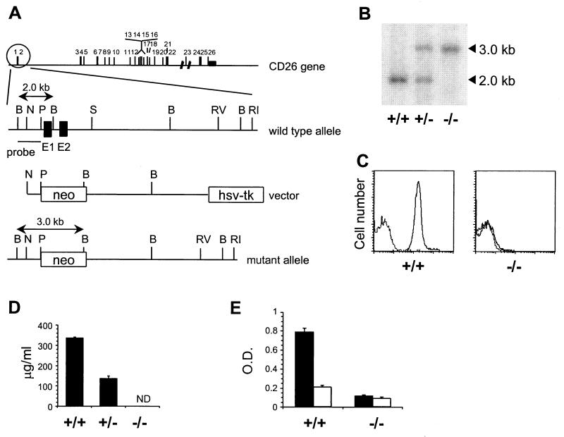
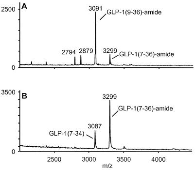
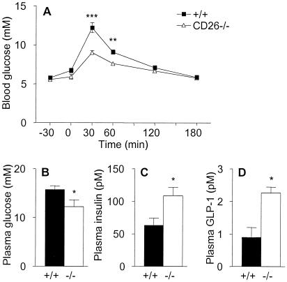
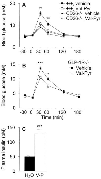
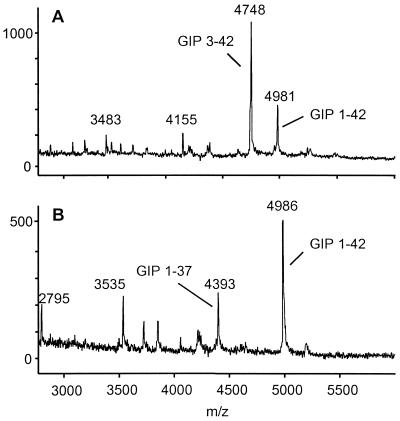
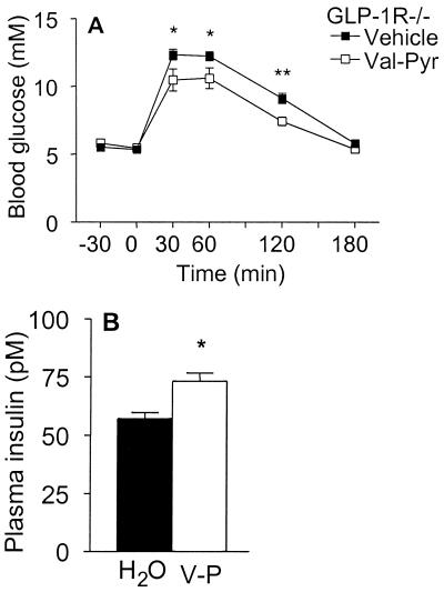
A subset of prolyl oligopeptidases, including dipeptidyl-peptidase IV (DPP IV or CD26, EC ), specifically cleave off N-terminal dipeptides from substrates having proline or alanine in amino acid position 2. This enzyme activity has been implicated in the regulation of the biological activity of multiple hormones and chemokines, including the insulinotropic peptides glucagon-like peptide 1 (GLP-1) and glucose-dependent insulinotropic polypeptide (GIP). Targeted inactivation of the CD26 gene yielded healthy mice that have normal blood glucose levels in the fasted state, but reduced glycemic excursion after a glucose challenge. Levels of glucose-stimulated circulating insulin and the intact insulinotropic form of GLP-1 are increased in CD26(-/-) mice. A pharmacological inhibitor of DPP IV enzymatic activity improved glucose tolerance in wild-type, but not in CD26(-/-), mice. This inhibitor also improved glucose tolerance in GLP-1 receptor(-/-) mice, indicating that CD26 contributes to blood glucose regulation by controlling the activity of GLP-1 as well as additional substrates. These data reveal a critical role for CD26 in physiological glucose homeostasis, and establish it as a potential target for therapy in type II diabetes.


Figures

Figure 1
Figure 1
Generation of CD26-deficient mice. (A) Targeting strategy. Exons in the CD26 gene are numbered. The wild-type CD26 allele, targeting construct, and expected mutant allele are shown with exon 1 and 2 of CD26 as filled boxes and the neo and TK genes as open boxes. Restriction sites: B, BglII; N, NcoI; P, PstI; S, SphI; RI, EcoRI, RV, EcoRV. (B) Southern blot containing BglII-digested DNA from CD26 wild-type (+/+), heterozygous (+/−), and homozygous mutant (−/−) mice hybridized with a 0.7-kb BglII–PstI probe specific for regions upstream of the targeting construct. (C) Flow cytometric analysis of CD26 expression at the surface of thymocytes from CD26 wild-type (+/+) and homozygous mutant (−/−) mice stained with anti-CD26 mAb followed by FITC-labeled anti-Ig (solid line) or with FITC-anti-Ig alone (dotted line). (D) Soluble CD26 protein in serum from CD26 wild-type (+/+), heterozygous (+/−), and homozygous mutant (−/−) mice measured by ELISA. ND, not detectable. (E) DPP IV enzyme activity in plasma from CD26 wild-type (+/+) and homozygous mutant (−/−) mice (n = 7). The DPP IV substrate Gly-Pro-pNA (pNA, p-nitroanilide) was incubated in plasma alone (black bars) or plasma containing 10 μM valine-pyrrolidide (open bars).
Figure 2
Figure 2
Lack of N-terminal degradation of GLP-1 in plasma from CD26−/− mice. Mass spectrometry analysis of GLP-1 degradation products after 4-h incubation of intact GLP-1-(7–36)-amide at 37°C in plasma from CD26 wild-type (A) or homozygous mutant (B) mice. The identity of peaks representing intact GLP-1-(7–36)-amide, N-terminally truncated GLP-1-(9–36)-amide, and C-terminally truncated GLP-1-(7–34) are indicated.
Figure 3
Figure 3
Enhanced oral glucose tolerance and increased levels of insulin and GLP-1 in CD26−/− mice. (A) Blood glucose concentrations measured at various times before and after oral administration of glucose (given at time 0) to female wild-type or CD26−/− mice, as indicated. n = 7. Similar data were obtained in a repeat experiment and in two experiments on groups of male mice. (B–D) Levels of glucose (B), insulin (C), and intact GLP-1-(7–36) (D) in plasma from male wild-type (closed bars) and CD26−/− (open bars) mice taken 15 min after oral glucose challenge. n = 8. *, P < 0.05; **, P < 0.01; ***, P < 0.001. Similar data were obtained in two additional experiments.
Figure 4
Figure 4
Specific pharmacological inhibition of CD26 enzymatic activity improves glucose tolerance, even in mice lacking GLP-1 signaling. (A) Female wild-type or CD26−/− mice were given vehicle (water) or 20.6 mg/kg valine-pyrrolidide (Val-Pyr) orally 30 min before oral glucose challenge. n = 8. Asterisks indicate level of significance of difference between wild-type mice given vehicle and valine-pyrrolidide. Similar data were obtained in another experiment with female mice and two experiments with male mice. Wild-type mice given valine-pyrrolidide exhibited significantly increased levels of insulin and intact GLP-1 at 15 min after the glucose challenge (data not shown). (B) Oral glucose tolerance test in male GLP-1R−/− mice given vehicle or 20.6 mg/kg valine-pyrrolidide 30 min before glucose challenge. n = 7–9. (C) Plasma insulin levels in GLP-1R−/− mice 15 min after oral glucose challenge. Mice were given vehicle (H2O) or valine-pyrrolidide (V-P) as indicated. n = 10–11. *, P < 0.05; **, P < 0.01; ***, P < 0.001.
Figure 5
Figure 5
CD26 is the major enzyme responsible for the N-terminal processing of GIP in serum. Mass spectrometry analysis of GIP degradation products after a 4-h incubation at 37°C in plasma from wild-type (A) or CD26−/− (B) mice. Peaks representing intact GIP-(1–42), N-terminally truncated GIP-(3–42), and C-terminally truncated GIP-(1–37) are indicated.
Figure 6
Figure 6
Inhibition of CD26 enzymatic activity accelerates clearance of glucose administered intraperitoneally in GLP-1R−/− mice. (A) Intraperitoneal glucose tolerance test in male GLP-1R−/− mice given vehicle (water) or valine-pyrrolidide at time −30 min and glucose at time 0. (B) Plasma insulin levels 15 min after glucose challenge in the animals shown in A, given vehicle (H2O) or valine-pyrrolidide (V-P), as indicated. n = 8–14. *, P < 0.05; **, P < 0.01.

References

    1. De Meester I, Korom S, Van Damme J, Scharpe S. Immunol Today. 1999;20:367–375. - PubMed
    1. Morimoto C, Schlossman S F. Immunol Rev. 1998;161:55–70. - PubMed
    1. Wesley U V, Albino A P, Tiwari S, Houghton A N. J Exp Med. 1999;190:311–322. - PMC - PubMed
    1. Kameoka J, Tanaka T, Nojima Y, Schlossman S F, Morimoto C. Science. 1993;261:466–469. - PubMed
    1. Marguet D, Bernard A M, Vivier I, Darmoul D, Naquet P, Pierres M. J Biol Chem. 1992;267:2200–2208. - PubMed

Publication types