

Internal packing of helical membrane proteins
- PMID: 10823938
- PMCID: PMC18513
- DOI: 10.1073/pnas.97.11.5796
Internal packing of helical membrane proteins
Abstract
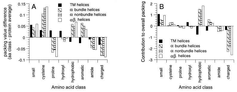
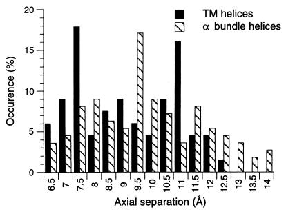
Helix packing is important in the folding, stability, and association of membrane proteins. Packing analysis of the helical portions of 7 integral membrane proteins and 37 soluble proteins show that the helices in membrane proteins have higher packing values (0.431) than in soluble proteins (0.405). The highest packing values in integral membrane proteins originate from small hydrophobic (G and A) and small hydroxyl-containing (S and T) amino acids, whereas in soluble proteins large hydrophobic and aromatic residues have the highest packing values. The highest packing values for membrane proteins are found in the transmembrane helix-helix interfaces. Glycine and alanine have the highest occurrence among the buried amino acids in membrane proteins, whereas leucine and alanine are the most common buried residue in soluble proteins. These observations are consistent with a shorter axial separation between helices in membrane proteins. The tight helix packing revealed in this analysis contributes to membrane protein stability and likely compensates for the lack of the hydrophobic effect as a driving force for helix-helix association in membranes.
Figures
References
-
- Richards F M. J Mol Biol. 1974;82:1–14. - PubMed
-
- Varadarajan R, Richards F M, Connelly P R. Curr Sci. 1990;59:819–824.
-
- Wang L, Veenstra D L, Radmer R J, Kollman P A. Proteins Struct Funct Genet. 1998;32:438–458. - PubMed
-
- DeDecker B S, O'Brien R, Fleming P J, Geiger J H, Jackson S P, Sigler P B. J Mol Biol. 1996;264:1072–1084. - PubMed
Publication types
MeSH terms
Substances
Grants and funding
LinkOut - more resources
Full Text Sources
Other Literature Sources
