Comparative Study
. 2000 May 23;97(11):5842-7.
doi: 10.1073/pnas.97.11.5842.

Structure of a two-domain chitotriosidase from Serratia marcescens at 1.9-A resolution

Affiliations
Comparative Study

Structure of a two-domain chitotriosidase from Serratia marcescens at 1.9-A resolution

D M van Aalten et al. Proc Natl Acad Sci U S A. .

Abstract

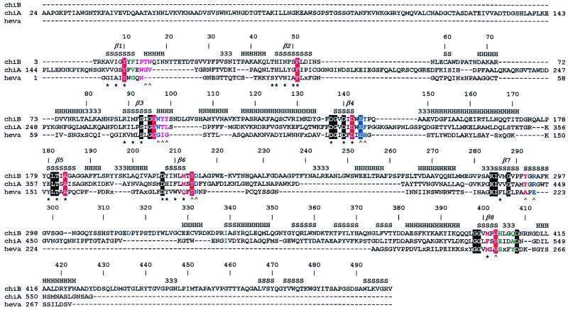
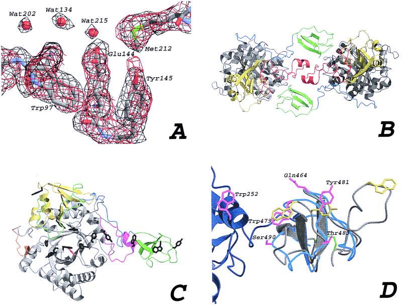
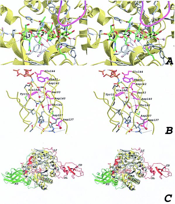
In this paper, we describe the structure of chitinase B from Serratia marcescens, which consists of a catalytic domain with a TIM-barrel fold and a 49-residue C-terminal chitin-binding domain. This chitinase is the first structure of a bacterial exochitinase, and it represents one of only a few examples of a glycosyl hydrolase structure having interacting catalytic and substrate-binding domains. The chitin-binding domain has exposed aromatic residues that contribute to a 55-A long continuous aromatic stretch extending into the active site. Binding of chitin oligomers is blocked beyond the -3 subsite, which explains why the enzyme has chitotriosidase activity and degrades the chitin chain from the nonreducing end. Comparison of the chitinase B structure with that of chitinase A explains why these enzymes act synergistically in the degradation of chitin.


Figures

Figure 1
Figure 1
Sequence alignment of chitinases. ChiB, chitinase B from S. marcescens; ChiA, chitinase A from S. marcescens; heva, hevamine from Hevea brasiliensis. An x in the sequences indicates a small insertion in hevamine. The secondary structure of ChiB is indicated at the top of the sequence alignment by H, α-helix; S, β-strand; 3, 310-helix. The TIM barrel β-strands are indicated by β1–8. Every 10th ChiB residue is labeled with its corresponding sequence number. Boxes indicate conserved residues. The color coding indicates the subsites of the chitotetraose model (see also Fig. 3A). Purple, −3; green, −2; red, −1; blue, +1. A ∧ indicates hydrogen bonding to an atom on the chitotetraose model, and a ★ indicates involvement of the side chain in the hydrogen-bonding network inside the TIM barrel core of ChiB. The catalytic Glu144 is part of the family 18 DxxDxDxE motif. The following definitions of loops and domains are used throughout the text: the porch loop (residues 14–27), the support loop (residues 233–262), the flexible loop (residues 315–325), the α/β-domain (residues 295–373) and the linker (residues 425–450), and the ChBD (residues 451–498).
Figure 2
Figure 2
(A) Comparison of experimental and final maps. An area around the active site residue Glu144 is drawn in a stick representation. A 1 σ contoured Fo,φMLPHARE map is shown in black, calculated by using the phases at the end of heavy atom refinement with MLPHARE. A 2Fo-Fccalc map is shown at the end of refinement with CNS, contoured at 1.4 σ (in red). (B) The two molecules in the asymmetric unit, color-coded to identify various regions. The TIM barrel (gray), the α/β-domain (yellow), the support loop (red), the linker (blue), and the ChBD (green). (C) ChiB, as in Fig. 2B, with the flexible loop covering the active site (green), the active site residue (red sticks), the porch loop (orange), and the exposed aromatic residues (black sticks). (D) Superposition of the ChBD of ChiB (blue ribbon) and the CeBD of endoglucanase Cel5 (gray ribbon). Most of the support loop of the catalytic domain of ChiB is shown as a dark-blue ribbon. Trp252 also is shown in magenta. The substrate-binding residues for the CeBD are shown in yellow, and the equivalent residues in the ChBD are shown in magenta. The disulfide bond between the termini of the CeBD is shown in green. Polar residues lining the path of aromatic residues in ChiB are shown in magenta. Labels correspond to the ChiB sequence. Note the almost exact overlap of the conserved β-strands.
Figure 3
Figure 3
(A) Stereo view of the active site with the modeled chitotetraose (same view as in Fig. 1C). The ChiB backbone is shown as a yellow ribbon. The modeled chitotetraose is shown in a stick representation, with the carbons colored green. Side chains within 5 Å of the chitotetraose are depicted by gray sticks, and also are indicated in Fig. 1. Possible hydrogen bonds are drawn as black dashed lines, and the residues involved are indicated in Fig. 1. The four water molecules that are predicted to be replaced by the substrate are shown as blue transparent spheres. The GlcNAc residues are labeled from −3 to +1, corresponding to their location with respect to the active site residue (15). The loop around residue 316, partially covering the active site, is shown in magenta. (B) Stereo view of the interior of the ChiB TIM barrel. The strands forming the TIM barrel are shown as a yellow ribbon. Side chains of residues lining the inside of the barrel are shown as sticks. Side chains conserved in ChiA, ChiB, and hevamine are colored magenta. Water molecules in the structure are shown as red spheres. Hydrogen bonds are shown as black dashed lines. Conserved residues are labeled according to the ChiB sequence. Part of the chitotetraose model is shown as sticks, with carbon atoms colored orange. (C) Stereo view of a superposition of ChiA and ChiB. Both structures are shown in a ribbon representation. ChiB is colored yellow, except for residues that correspond to insertions in ChiB with respect to ChiA, which are colored red. ChiA is colored gray except for residues that correspond to insertions in ChiA with respect to ChiB, which are colored green. Some insertions are indicated with two-letter labels. AA, active site covering loop in ChiA; AB, active site covering loop in ChiB; CD, ChBD in ChiB; DL, ChBD support loop in ChiB; FD, fibronectin domain in ChiA; LI, linker in ChiB; PO, porch loop in ChiB.

References

    1. Fuchs R L, McPherson S A, Drahos D J. Appl Environ Microbiol. 1986;51:504–509. - PMC - PubMed
    1. Perrakis A, Tews I, Dauter Z, Oppenheim A B, Chet I, Wilson K S, Vorgias C E. Structure (London) 1994;2:1169–1180. - PubMed
    1. Tews I, Perrakis A, Oppenheim A, Dauter Z, Wilson K S, Vorgias C E. Nat Struct Biol. 1996;3:638–648. - PubMed
    1. Watanabe T, Kimura K, Sumiya T, Nikaidou M, Suzuki K, Suzuki M, Taiyoji M, Ferrer S, Regue M. J Bacteriol. 1997;179:7111–7117. - PMC - PubMed
    1. Perrakis A, Ouzonis C, Wilson K S. Folding Des. 1997;2:291–294. - PubMed

Publication types

MeSH terms

Associated data

LinkOut - more resources