. 2000 May 23;97(11):5930-5.
doi: 10.1073/pnas.97.11.5930.

Human vascular endothelial cells contain membrane binding sites for estradiol, which mediate rapid intracellular signaling

Affiliations

Human vascular endothelial cells contain membrane binding sites for estradiol, which mediate rapid intracellular signaling

K S Russell et al. Proc Natl Acad Sci U S A. .

Abstract

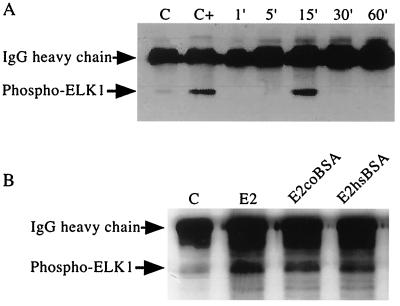
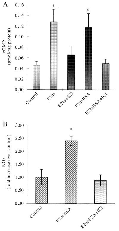
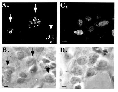
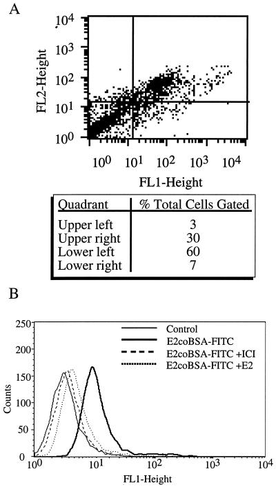
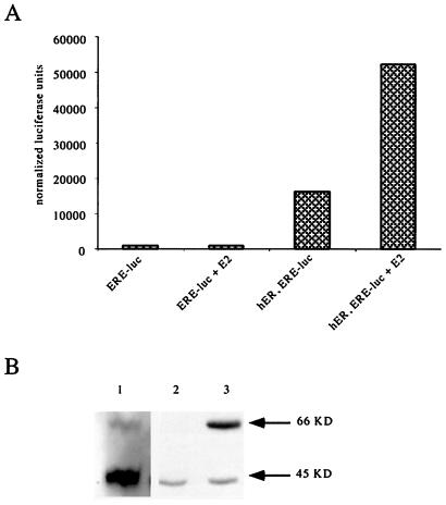
Estrogen induces both rapid and delayed effects on the cardiovascular system. The early effects take place within minutes (e.g., changes in vasomotor tone) and are mediated through rapid intracellular signaling pathways; whereas the delayed effects (e.g., remodeling or lipid alterations) require hours to days to occur and require transcriptional effects with subsequent modulation of protein expression. To study the acute effects of 17beta-estradiol (E2) treatment on vascular function, we have investigated the rapid (on the order of minutes) effects of E2 treatment on intracellular signaling in human endothelial cells (EC). Our previous data have shown that E2 induces rapid release of NO from and activation of guanylate cyclase in human EC. In this study, we demonstrate that E2 also activates mitogen-activated protein kinase (extracellular signal-related kinase) signaling within minutes in EC. We hypothesized that this effect might be mediated by estrogen receptors (ER) localized to the cell surface. Our data show that membrane-impermeant forms of E2 also activate EC mitogen-activated protein kinase as well as stimulate cGMP production and NO release. The ER antagonist ICI 182,780 blocks this effect. Using confocal microscopy and flow cytometric analysis, we demonstrate that EC contain surface binding sites for E2, detectable by cell-impermeant ligand binding and equally with an anti-ERalpha antibody. Immunoreactive bands of 66 and 45 kDa are detectable with an anti-ERalpha mAb in human EC, and their individual presence correlates functionally with E2-stimulated genomic and rapid nongenomic responses, respectively. Membrane ERs may provide key molecular switches in these novel, rapid signaling pathways induced by E2 in EC.


Figures

Figure 1
Figure 1
HUVEC MAPK activation by free and membrane-impermeant forms of estrogen. Serum-deprived HUVEC monolayers were E2 (50 ng/ml)-treated for the indicated times (A) or treated for 15 min with either E2 (50 ng/ml), E2coBSA (395 ng/ml, equivalent to 50 ng/ml of E2), or E2hsBSA (308 ng/ml, equivalent to 50 ng/ml of E2) (B). Subsequently, activated ERK1/2, immunoprecipitated from cell lysates, was used in an in vitro kinase assay with ELK-1 peptide substrate. Kinase reaction samples were immunoblotted with a phospho-specific anti-ELK-1 antibody. Controls were treated with 5% BSA containing media alone (C) or the addition of purified activated MAPK to the kinase reaction (C+).
Figure 2
Figure 2
cGMP and NOx production induced by membrane-impermeant E2. (A) HUVEC monolayers were treated with E2hs (equivalent to 10 ng/ml of E2) or E2hsBSA (equivalent to 10 ng/ml E2) in the presence or absence of the ER antagonist ICI 182,780 (10 μM) for 20 min at 37°C, after which cGMP levels were measured by RIA. (B) EA.hy926 monolayers were treated with E2coBSA (equivalent to 10 ng/ml of E2) in the absence or presence of ICI 182,780 for 30 min at 37°C, after which the amount of NOx released into the medium was determined by NO-specific chemiluminescence. *P < 0.01 vs. control.
Figure 3
Figure 3
Effect of EC permeabilization on immunofluorescent staining with membrane-impermeant estradiol. HUVEC monolayers grown on collagen-coated glass coverslips were stained on ice without fixation (A and B) or after fixation and permeabilization (C and D) by incubation with E2coBSA-FITC (1:100) in PBS with 5% BSA. Confocal immunofluorescence (A and C) or corresponding phase-contrast (B and D, respectively) micrographs are shown. Arrows denote intact cells staining with E2coBSA-FITC (A) and corresponding cells shown by phase microscopy (C). The photomicrographs shown are representative of multiple fields examined in three experiments (bar, 2.5 μM).
Figure 4
Figure 4
FACS analysis with membrane-impermeant E2 and anti-ER antibody. (A) HUVEC were harvested by nonenzymatic cell dissociation and single-cell suspensions of nonpermeabilized cells stained with E2coBSA-FITC and anti-ERα mAb 1D5 plus GAM-PE in the presence of 5% BSA. Background gating of cells stained with FITC and GAM-PE alone was performed as a negative control (99% of cells within left lower quadrant, data not shown). Five thousand cells were analyzed per sample. FL1-Height: FITC fluorescence relative to gated background; FL2-Height: PE fluorescence relative to gated background. Statistical analysis of quadrants is shown. (B) EA.hy926 monolayer cells were harvested and single-cell suspensions of nonpermeabilized cells stained with E2coBSA-FITC in the presence of 5% BSA. Indicated samples were treated on ice with ICI 182,780 or a 20-fold excess of E2 for 1 h before E2coBSA-FITC staining. Five thousand cytometer-acquired events were analyzed per sample.
Figure 5
Figure 5
Structure-function correlation between different forms of estrogen receptors. (A) EA.hy926 cells were transiently cotransfected with ERE-luciferase (ERE-luc) and cytomegalovirus β-galactosidase reporter constructs, with or without a full-length ERα expression construct (hER). Twenty-four hours posttransfection and 48 h postestrogen deprivation, transfected monolayers were stimulated with E2 or vehicle control, and luciferase activity was measured in cell extracts after 24 h. Counts represent β-galactosidase-normalized luciferase units. (B) Western blot analysis for ERα (immunoblotting with anti-C-terminal ERα mAb F-10) was performed on total cell lysates obtained from HUVEC (lane 1), EA.hy926 cells (lane 2), and EA.hy926 cells transiently transfected with hER (lane 3). Lane 2 cells are representative of those used in columns 1 and 2 experimental samples (A, above) and lane 3 cells are representative of those used in columns 3 and 4 (A, above).

References

    1. Caulin-Glaser T, Garcia-Cardena G, Sarrel P, Sessa W C, Bender J R. Circ Res. 1997;81:885–892. - PubMed
    1. Blackmore P F, Neulen J, Lattanzio F, Beebe S J. J Biol Chem. 1991;266:18655–18659. - PubMed
    1. Szego C M, Pietras R J. Nature (London) 1985;317:88–89. - PubMed
    1. Pietras R J, Szego C M. Nature (London) 1977;265:69–72. - PubMed
    1. Razandi M, Pedram A, Greene G L, Levin E R. Mol Endocrinol. 1999;13:307–319. - PubMed

Publication types

MeSH terms

LinkOut - more resources