. 2000 May 23;97(11):6013-8.
doi: 10.1073/pnas.97.11.6013.

Cholesterol-dependent clustering of IL-2Ralpha and its colocalization with HLA and CD48 on T lymphoma cells suggest their functional association with lipid rafts

Affiliations

Cholesterol-dependent clustering of IL-2Ralpha and its colocalization with HLA and CD48 on T lymphoma cells suggest their functional association with lipid rafts

G Vereb et al. Proc Natl Acad Sci U S A. .

Abstract

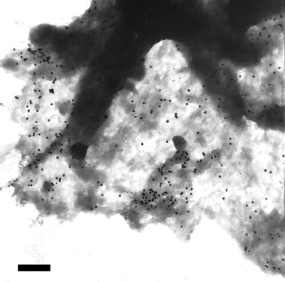
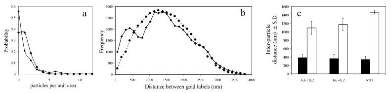
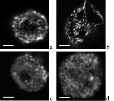
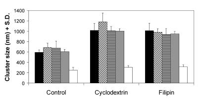
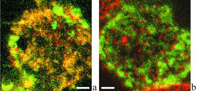
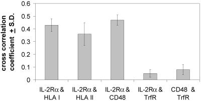
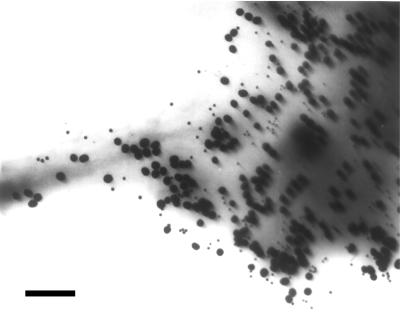
Immunogold staining and electron microscopy show that IL-2 receptor alpha-subunits exhibit nonrandom surface distribution on human T lymphoma cells. Analysis of interparticle distances reveals that this clustering on the scale of a few hundred nanometers is independent of the presence of IL-2 and of the expression of the IL-2R beta-subunit. Clustering of IL-2Ralpha is confirmed by confocal microscopy, yielding the same average cluster size, approximately 600-800 nm, as electron microscopy. HLA class I and II and CD48 molecules also form clusters of the same size. Disruption of cholesterol-rich lipid rafts with filipin or depletion of membrane cholesterol with methyl-beta-cyclodextrin results in the blurring of cluster boundaries and an apparent dispersion of clusters for all four proteins. Interestingly, the transferrin receptor, which is thought to be located outside lipid rafts, exhibits clusters that are only 300 nm in size and are less affected by modifying the membrane cholesterol content. Furthermore, transferrin receptor clusters hardly colocalize with IL-2Ralpha, HLA, and CD48 molecules (crosscorrelation coefficient is 0.05), whereas IL-2Ralpha colocalizes with both HLA and CD48 (crosscorrelation coefficient is between 0.37 and 0.46). This coclustering is confirmed by electron microscopy. The submicron clusters of IL-2Ralpha chains and their coclustering with HLA and CD48, presumably associated with lipid rafts, could underlie the efficiency of signaling in lymphoid cells.


Figures

Figure 1
Figure 1
Nonrandom distribution of IL-2Rα on Kit 225 K6 cells revealed by colloidal gold labels. Kit 225 K6 cells were labeled with anti-Tac as primary antibody and then with 30 nm colloidal gold conjugated to the secondary antibodies. A representative electron micrograph of the periphery of a cell is shown. The distribution of gold labels appears to be nonrandom. Bar = 470 nm.
Figure 2
Figure 2
Quantitative analysis of the distribution of gold labels on IL-2Rα-subunits. (a) Gold labels shown in Fig. 1 were counted (n = 406), and the unit area was defined such that the expected value of gold labels per unit area was one. The image was divided into equal squares of one unit area each and the actual distribution of labels among the squares determined. The probability distribution of the particle density per unit area is plotted for the actual finding (♦) and compared to a Poisson distribution with parameter λ = 1(●). (b) The coordinates of all labels in Fig. 1 were determined, and the distribution of all interparticle distances was plotted (♦). A model distribution was also generated assuming Poissonian statistics (dashed line, ●). In contrast to the single peak of the expected random distribution, the measured distribution has two peaks. The first peak, around 400 nm, represents the characteristic distance of gold labels within clusters. (c) Characteristic distances for gold labels determined as in b are plotted for Kit 225 K6 (K6 + IL2), IL-2-starved Kit 225 K6 (K6-IL2), and MT-1 cells. Characteristic distances within clusters are represented by filled columns, and average distances within the whole sample area are shown with open columns. Data are mean ± SD from six independent experiments.
Figure 3
Figure 3
Confocal laser-scanning microscopy of Cy3-labeled IL-2Rα and TrfR. Kit 225 K6 cells were labeled with Cy3-conjugated αTac Fab against the IL-2Rα-subunit (a, c, and d) or XR-conjugated MEM-75 against the TrfR (b). Cells in c and d were treated with filipin and methyl-β-cyclodextrin, respectively. Confocal slices of 0.6 μm thickness were obtained. Surface fluorescence distribution was reconstructed from z directional projection of image slices. Bar = 4 μm. A patchy receptor distribution can be observed with clusters of 200- to 1,200-nm diameter depending on the type of receptor and the treatment.
Figure 4
Figure 4
Cluster sizes of IL-2Rα, HLA class I and II, CD48, and TrfR and their modulation by membrane cholesterol content. Cluster sizes on Kit 225 K6 cells determined from the angle-averaged autocorrelation function are presented for IL-2Rα (filled columns), HLA class I (crosshatched columns), and class II (striped columns), CD48 (gray columns), and TrfR (open columns). The effect of modulating the cholesterol content of the membrane is also displayed: with the exception of TrfR, all receptor clusters exhibit a significant increase of cluster size on both cholesterol depletion by cyclodextrin or in situ complexation of cholesterol by filipin. (n > 9, from three independent experiments).
Figure 5
Figure 5
IL-2Rα exhibits submicron-scale colocalization with MHC II but not with TrfR. A representative confocal fluorescence image of the colocalization of IL-2Rα and HLA class II is shown in a. IL-2Rα and HLA class II are labeled with XF (green) and XR (red), respectively. Because of the high degree of colocalization, many pixels appear orange when the two channels are fused. b demonstrates that IL-2Rα (green) and TrfR (red) codetected in a similar experiment are mostly localized at different areas of the plasma membrane. Bar = 2 μm.
Figure 6
Figure 6
IL-2Rα staining crosscorrelates with MHC glycoproteins and CD48 but not with TrfR. Kit 225 K6 cells were double labeled with pairs of antibodies against IL-2Rα, HLA class I and II, CD48, and TrfR. The crosscorrelation coefficient is measured for the following receptor pairs: IL-2Rα and HLA class I, IL-2Rα and HLA class II, IL-2Rα and CD48, IL-2Rα and TrfR, and CD48 and TrfR. While IL-2Rα colocalizes with HLA class I and II and CD48, a raft marker, neither IL-2Rα nor CD48 colocalizes with TrfR (n = 7).
Figure 7
Figure 7
Electron microscopy confirms the partial coclustering of IL-2Rα and MHC molecules. Immunogold labeling followed the sequence: αTac Fab—10 nm AuroGamig—blocking by αTac Fab—W6/32 whole antibody—30 nm AuroGamig (anti-Fc). Electron microscopy shows that the selective labels against the IL-2Rα and the MHC are partially, although not completely, colocalized, thus confirming the confocal microscopic data (Bar = 200 nm).

References

    1. Waldmann T A. Science. 1986;232:727–732. - PubMed
    1. Waldmann T A. J Biol Chem. 1991;266:2681–2684. - PubMed
    1. Nakamura Y, Russell S M, Mess S A, Friedmann M, Erdos M, Francois C, Jacques Y, Adelstein S, Leonard W J. Nature (London) 1994;369:330–333. - PubMed
    1. Damjanovich S, Bene L, Matkó J, Alileche A, Goldman C K, Sharrow S, Waldmann T A. Proc Natl Acad Sci USA. 1997;94:13134–13139. - PMC - PubMed
    1. Damjanovich S, Gáspár R, Jr, Pieri C. Q Rev Biophys. 1997;30:67–106. - PubMed

Publication types