. 2000 May 23;97(11):6043-8.
doi: 10.1073/pnas.97.11.6043.

Inflammation in the developing human intestine: A possible pathophysiologic contribution to necrotizing enterocolitis

Affiliations

Inflammation in the developing human intestine: A possible pathophysiologic contribution to necrotizing enterocolitis

N N Nanthakumar et al. Proc Natl Acad Sci U S A. .

Abstract

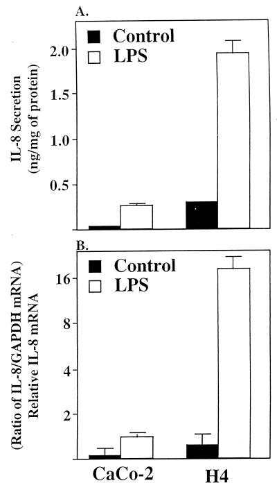
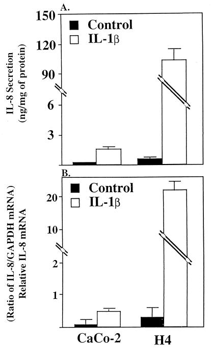
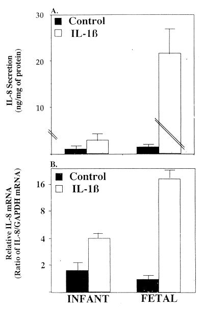
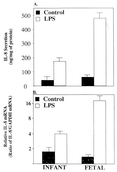
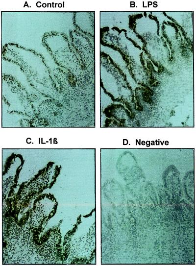
Necrotizing enterocolitis (NEC), a major cause of morbidity and mortality in premature infants, occurs after the introduction of oral feedings in conjunction with initial bacterial colonization of the gut and is hypothesized to be due to an immature (inappropriate) enterocyte response to bacterial stimuli. To test this hypothesis, we compared the enterocyte IL-8 response to inflammatory stimuli [lipopolysaccharide (LPS) and IL-1beta] in immature vs. mature human small intestine. Initial in vitro studies comparing confluent Caco-2 cells, a model for mature human enterocytes, with a primary human fetal intestinal cell line (H4 cells) demonstrated that after inflammatory stimulation fetal cells secreted more IL-8 (LPS, 8-fold; IL-1beta, 20-fold) than Caco-2 cells. IL-8 mRNA activity in fetal compared to Caco-2 cells was proportionately increased by the same magnitude with both stimuli. To validate the in vitro observations, small intestinal organ cultures from fetuses vs. older children were exposed to LPS and IL-1beta. Again in human organ cultures from fetuses compared to older children, IL-8 secretion was greater (LPS, 2.5-fold; IL-1beta, 200-fold) and mRNA activity after stimulation was comparably higher, suggesting that increased transcription of the IL-8 gene may account for the excessive response. Using immunohistochemical staining to identify the cellular source of IL-8, activity was noted predominantly in villous and crypt epithelium but also in a few immunoresponsive lymphoid cells. The observation that immature human enterocytes react with excessive pro-inflammatory cytokine production after inflammatory stimulation may help in part explain why prematures exposed to initial colonizing bacteria develop necrotizing enterocolitis.


Figures

Figure 1
Figure 1
Depiction of IL-8 secretion (A) and IL-8 mRNA induction (B) in confluent Caco-2 and fetal human enterocytes (H4 cells) in response to LPS (50 μg/ml) or media alone as a control. Secreted IL-8 is expressed as ng/mg of total cellular protein. IL-8 mRNA was quatitated by densitometry and normalized to the relative level of GAPDH mRNA. Results are given as means ± SEM. The number of independent experiments for each data point ranged from four to five and an absence of an error bar indicates the SEM is smaller than the symbol.
Figure 2
Figure 2
Depiction of IL-8 secretion (A) and IL-8 mRNA induction (B) in confluent Caco-2 and fetal human enterocytes (H4 cells) in response to IL-1β (1 ng/ml) or media alone as a control. Secreted IL-8 is expressed as ng/mg of total cellular protein. IL-8 mRNA was quatitated by densitometry and normalized to the relative level of GAPDH mRNA. The results are given as means ± SEM. The number of independent experiments for each data point ranged from four to five and as absence of error bar indicates the SEM is smaller than the symbol.
Figure 3
Figure 3
Depiction of IL-8 secretion (A) and IL-8 mRNA induction (B) in infant and fetal intestinal organ culture in response to LPS (50 μg/ml) or media alone as a control. Secreted IL-8 is expressed as ng/mg of total tissue protein. IL-8 mRNA was quatitated by densitometry and normalized to the relative level of GAPDH mRNA. The results are given as means ± SEM. The number of independent experiments for each data point ranged from three to four.
Figure 4
Figure 4
Depiction of IL-8 secretion (A) and IL-8 mRNA induction (B) in infant and fetal intestinal organ culture in response to IL-1β (1 ng/ml) or media alone as a control. Secreted IL-8 is expressed as ng/mg of total tissue protein. IL-8 mRNA was quatitated by densitometry and normalized to the relative level of GAPDH mRNA. The results are given as means ± SEM. The number of independent experiments for each data point ranged from three to four.
Figure 5
Figure 5
This figure depicts IL-8 immunoreactivity in microscopic sections from human organ cultures after treatment with control media (A), LPS (B), or IL-1β (C). An additional microscopic section from a LPS-treated organ culture section stained with a class specific control antibody is shown in D. It should be noted that crypt and differentiated villus epithelial compartments are strongly positive for IL-8 with a few lymphoid cells in the lamina propria showing a reaction whereas no immunoreactivity is noted in D (AD, ×50X, Toluidine Blue).

References

    1. Kliegman R M, Walker W A, Yolken R H. Pediatr Res. 1983;34:701. - PubMed
    1. Neu J. Pediatr Clin North Am. 1996;43:409. - PMC - PubMed
    1. MacKendrick W, Caplan M. Pediatr Clin North Am. 1993;40:1047. - PubMed
    1. Foglia R P. Curr Prob Surg. 1994;32:757–823. - PubMed
    1. Stoll B J. Clin Perinatol. 1994;21:205–216. - PMC - PubMed

Publication types

MeSH terms

LinkOut - more resources