Comparative Study
. 2000 May 23;97(11):6144-9.
doi: 10.1073/pnas.97.11.6144.

Classification of fusiform neocortical interneurons based on unsupervised clustering

Affiliations
Comparative Study

Classification of fusiform neocortical interneurons based on unsupervised clustering

B Cauli et al. Proc Natl Acad Sci U S A. .

Abstract

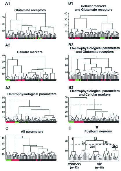
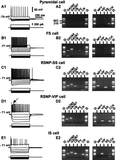
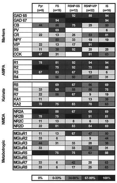
A classification of fusiform neocortical interneurons (n = 60) was performed with an unsupervised cluster analysis based on the comparison of multiple electrophysiological and molecular parameters studied by patch-clamp and single-cell multiplex reverse transcription-PCR in rat neocortical acute slices. The multiplex reverse transcription-PCR protocol was designed to detect simultaneously the expression of GAD65, GAD67, calbindin, parvalbumin, calretinin, neuropeptide Y, vasoactive intestinal peptide (VIP), somatostatin (SS), cholecystokinin, alpha-amino-3-hydroxy-5-methyl-4-isoxazolepropionic acid, kainate, N-methyl-d-aspartate, and metabotropic glutamate receptor subtypes. Three groups of fusiform interneurons with distinctive features were disclosed by the cluster analysis. The first type of fusiform neuron (n = 12), termed regular spiking nonpyramidal (RSNP)-SS cluster, was characterized by a firing pattern of RSNP cells and by a high occurrence of SS. The second type of fusiform neuron (n = 32), termed RSNP-VIP cluster, predominantly expressed VIP and also showed firing properties of RSNP neurons with accommodation profiles different from those of RSNP-SS cells. Finally, the last type of fusiform neuron (n = 16) contained a majority of irregular spiking-VIPergic neurons. In addition, the analysis of glutamate receptors revealed cell-type-specific expression profiles. This study shows that combinations of multiple independent criteria define distinct neocortical populations of interneurons potentially involved in specific functions.


Figures

Figure 1
Figure 1
Comparison of different cluster analyses applied to neocortical fusiform interneurons (black boxes, n = 60) as well as to pyramidal (green boxes, n = 9) and FS (red boxes, n = 16) cells taken as controls. For each diagram, the x axis represents the individuals, and the y axis represents the average within-cluster linkage distance. Dotted lines in B3 and D indicate the limits between clusters as suggested by the Thorndike procedure (see Materials and Methods). (A) Analyses based on the expression profiles of glutamate receptors (A1), of cellular markers (A2), or on electrophysiological properties (A3). (B and C) Analyses based on the combination of two of three sets of parameters (B1, B2, and B3) or on all parameters (C). Note that the control pyramidal and FS cells were segregated only by the combination of electrophysiological parameters and of cellular markers and that the expression of glutamate receptors blurs the classification. (D) Cluster analysis with the same parameters as in B3 restricted to the fusiform cell population. This analysis disclosed three groups of fusiform RSNP cells mainly expressing SS (RSNP-SS; branch labeled 1; n = 12) or mainly expressing VIP (branch labeled 2; n = 48). Within the group of VIP expressing cells, most of neurons of branch 2a were IS interneurons, and the majority of cells in branch 2b were RSNP cells. Within branch 2b, two subpopulations of RSNP-VIP (branches 2b1 and 2b2) were suggested by the Thorndike procedure.
Figure 2
Figure 2
Electrophysiological and biochemical analysis of five types of neocortical neurons. (Left) The firing properties of the five neurons. Membrane potentials were adjusted as indicated to the left of the recordings. (Right) The molecular analysis of the cells shown at Left. PCR products were resolved by agarose gel electrophoresis with Φ×174/HaeIII as a molecular mass marker. (A) Pyramidal neurons. (A1) In response to depolarizing current step (300 pA), the pyramidal cell fired action potentials with a marked frequency adaptation (Upper). (A2) RT-mPCR analysis showing only the expression of CCK mRNA. The faint band (≈800 bp) observed in the SS lane corresponds to the genomic amplification of SS. (B) FS interneuron. (B1) Note the high frequency, nonadapting discharge of fast action potentials with large hyperpolarized afterpotentials (Upper; 150 pA). (B2) The same FS cell expressed GAD65, GAD67, and PV. (C) RSNP-SS cell. (C1) Injection of a depolarizing current step produced an accommodating discharge of action potentials (Upper; 250 pA). Note the progressive reduction in the amplitude of the action potentials during the discharge. (C2) GAD65, GAD67, CB, NPY, and SS were detected in this neuron. (D) RSNP-VIP neuron. (D1) Injection of depolarizing current (Upper; 200 pA) elicited the firing of action potentials with a marked frequency adaptation. Note the augmentation after a marked reduction of spike amplitude (arrow). (D2) Agarose gel showing the expression of GAD65, GAD67, VIP, and CCK mRNAs. (E) IS interneuron. (E1) In response to depolarizing current, the neuron discharged an initial burst of action potentials followed by irregularly emitted action potentials. (E2) The agarose gel shows the expression of GAD65, GAD67, CB, CR, VIP, and CCK mRNAs.
Figure 3
Figure 3
Expression patterns of molecular markers and glutamate receptors in different classes of neocortical neurons. The percentage of neurons expressing a given mRNA is represented by a gray scale, as indicated on the bottom of the figure. Pyramidal cells (Pyr) did not express GADs in contrast to the other classes of neurons. FS cells consistently expressed PV and NPY. The majority of RSNP-SS cells expressed SS, whereas most of RSNP-VIP and IS cells expressed VIP. The distribution of α-amino-3-hydroxy-5-methyl-4-isoxazolepropionic acid (AMPA) receptor subunits revealed that the five classes of neurons frequently expressed GluR1 and GluR2. GluR3 was very rare in RSNP-VIP and IS. Pyramidal cells lacked GluR4 subunits. The distribution of kainate receptor subunits showed that KA2 subunit was the most frequently observed subunit. GluR5 subunit was not expressed in pyramidal cells. Distribution of N-methyl-d-aspartate (NMDA) subunits indicated that NR2A and NR2B were detected frequently in the five classes of cells. NR2D was more frequent in interneurons than in pyramidal cells. The distribution of mGluRs showed that mGluR1 was frequent in fusiform cells. mGluR5 was the most frequently expressed mGluR. Group III mGluRs were rare in FS cells.

References

    1. Baughman R W, Gilbert C D. J Neurosci. 1981;1:427–439. - PMC - PubMed
    1. Houser C R, Hendry S H, Jones E G, Vaughn J E. J Neurocytol. 1983;12:617–638. - PubMed
    1. Ramón y Cajal S. General Structure of the Cerebral Cortex. 1995. trans. Swanson, N. & Swanson, R. W. (Oxford Univ. Press, New York), pp. 429–492.
    1. Celio M R. Science. 1986;231:995–997. - PubMed
    1. van Brederode J F, Helliesen M K, Hendrickson A E. Neuroscience. 1991;44:157–171. - PubMed

Publication types

LinkOut - more resources