Comparative Study
. 2000 May 23;97(11):6185-90.
doi: 10.1073/pnas.97.11.6185.

Opposing effects of protein kinase C and protein kinase A on metabotropic glutamate receptor signaling: selective desensitization of the inositol trisphosphate/Ca2+ pathway by phosphorylation of the receptor-G protein-coupling domain

Affiliations
Comparative Study

Opposing effects of protein kinase C and protein kinase A on metabotropic glutamate receptor signaling: selective desensitization of the inositol trisphosphate/Ca2+ pathway by phosphorylation of the receptor-G protein-coupling domain

A Francesconi et al. Proc Natl Acad Sci U S A. .

Abstract

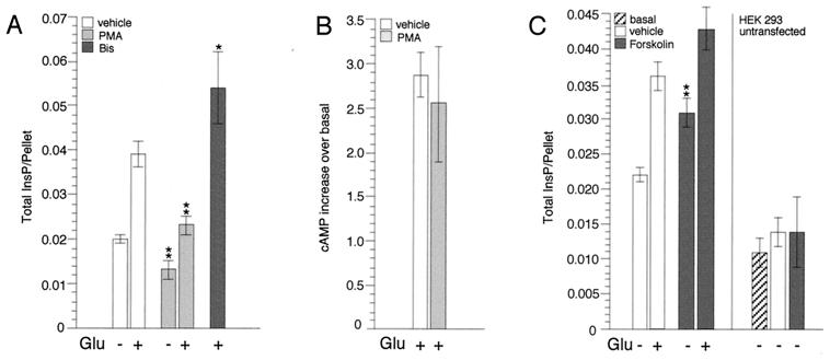
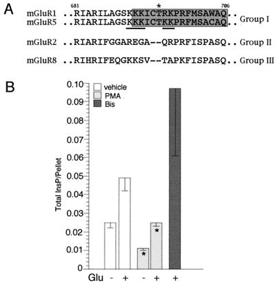
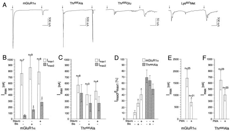
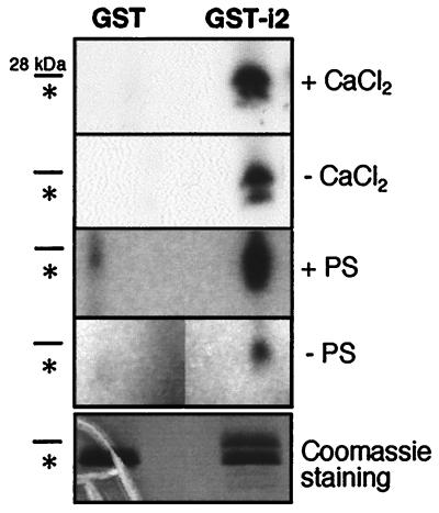
Signaling by the metabotropic glutamate receptor 1alpha (mGluR1alpha) can lead to the accumulation of inositol 1,4, 5-trisphosphate (InsP(3)) and cAMP and to the modulation of K(+) and Ca(2+) channel opening. At present, very little is known about how these different actions are integrated and eventually turned off. Unraveling the molecular mechanisms underlying these functions is crucial for understanding mGluR-mediated regulation of synaptic transmission. It has been shown that receptor-induced activation of the InsP(3) pathway is subject to feedback inhibition mediated by protein kinase C (PKC). In this study, we provide evidence for a differential regulation by PKC and protein kinase A of two distinct mGluR1alpha-dependent signaling pathways. PKC activation selectively inhibits agonist-dependent stimulation of the InsP(3) pathway but does not affect receptor signaling via cAMP. In contrast, protein kinase A potentiates agonist-independent signaling of the receptor via InsP(3). Furthermore, we demonstrate that the selectivity of PKC action on receptor signaling rests on phosphorylation of a threonine residue located in the G protein-interacting domain of the receptor. Modification at Thr(695) selectively disrupts mGluR1alpha-G(q/11) interaction without affecting signaling through G(s). Together, these data provide insight on the mechanisms by which selective down-regulation of a specific receptor-dependent signaling pathway can be achieved and on how cross-talk between different second messenger cascades may contribute to fine-tune short- and long-term receptor activity.


Figures

Figure 1
Figure 1
PKC and PKA regulate mGluR1α signaling. (A) Effect of PMA and bisindolylmaleimide I (Bis) on mGluR1α-dependent InsP accumulation. For each experiment, HEK 293 cells were transfected with 10 μg of mGluR1α and 10 μg of carrier DNA and seeded into six wells of a 24-well cluster. Stimulation was carried out in Hanks' saline solution (containing divalents and glucose) after clearing glutamate from the media with glutamic-pyruvic transaminase/pyruvate (9). The cells were treated with either Me2SO (vehicle) or test reagents and then stimulated with Glu. Results represent means ± SEM of duplicate or triplicate determinations obtained from at least two independent experiments (*, P ≤ 0.05; **, P ≤ 0.01; two-tailed t test). (B) Effect of PMA on mGluR1α-dependent cAMP accumulation. HEK 293 cells were transfected with 5 μg of mGluR1α along with 5 μg of Gs and 10 μg of carrier DNA and seeded into four wells of a 24-well cluster. The cells were preincubated with either Me2SO or PMA for 30 min and then stimulated with Glu for 20 min in the continued presence of the test reagents. Results represent means ± SEM of duplicate determinations obtained from three independent experiments. (C) Effect of forskolin on mGluR1α-dependent InsP accumulation. Cell transfection and treatment were conducted as described in A; in control experiments, 4 × 10⁵ untransfected cells were plated for each well. Results represent means ± SEM of triplicate determinations obtained from three independent experiments; for untransfected cells, results shown are means ± SD of triplicate determinations, representative of two independent experiments.
Figure 2
Figure 2
(A) Sequence alignment of the second intracellular loop of mGluRs; the region involved in selective coupling of mGluR1α with Gq/11 is boxed in gray. An asterisk highlights Thr695, which we propose to be substrate for PKC-mediated phosphorylation. Basic amino acid residues which can constitute the consensus for PKC are underlined. (B) Effect of PMA and bisindolylmaleimide I (Bis) on the Thr695Ala mutant receptor activity. Cell transfection and treatment were conducted as described for the wild-type receptor (Fig. 1A). Results represent means ± SEM of duplicate or triplicate determinations obtained from at least two independent experiments (*, P ≤ 0.05; two-tailed t test).
Figure 3
Figure 3
Rapid desensitization of mGluR1α depends on the phosphorylation state of Thr695. (A) Representative tracings of currents generated by wild-type and mutant receptors expressed in oocytes upon repeated application of Glu (1 mM); arrows indicate beginning of Glu application which lasted for 1 min. Time scale: 1 min. (B–F) PKC dependence of wild-type and mutant receptor responses. Injected oocytes were incubated with either staurosporin (100 nM) or bisindolylmaleimide I (2 μM) or PMA (100 nM) before recordings. Results are means ± SEM of values obtained from two independent experiments (*, P ≤ 0.05; **, P ≤ 0.01; two-tailed t test).
Figure 4
Figure 4
Thr695 is phosphorylated in vitro. Recombinant fusion protein (GST-i2) and GST were used as substrate for phosphorylation by PKC purified from brain. Data shown are representative of three independent experiments. To verify for the specificity of the phosphorylation, the effects of lipids [phosphatidyl serine (PS)] and calcium on enzyme activity were tested. GST-i2 predicted molecular mass is 28 kDa; GST alone is 26 kDa and its position on the gel is indicated by an asterisk. The gels were exposed for autoradiography at −70°C for 12 h (with or without CaCl2) and 8 h (with or without PS), respectively.

References

    1. Pin J-P, Duvoisin R M. Neuropharmacology. 1995;34:1–26. - PubMed
    1. Conn P J, Pin J-P. Annu Rev Pharmacol Toxicol. 1997;37:205–237. - PubMed
    1. Nakanishi S. Neuron. 1994;13:1031–1037. - PubMed
    1. Gudermann, N. C., Gahwiler, B. H. & Schultz, G. (1996) Annu. Rev. Pharmacol. Toxicol.429–459. - PubMed
    1. Clapham D E, Neer E J. Annu Rev Pharmacol Toxicol. 1997;37:167–203. - PubMed

Publication types

MeSH terms

LinkOut - more resources