Comparative Study
. 2000 May 23;97(11):6224-9.
doi: 10.1073/pnas.97.11.6224.

A calcium-dependent protein kinase can inhibit a calmodulin-stimulated Ca2+ pump (ACA2) located in the endoplasmic reticulum of Arabidopsis

Collaborators, Affiliations
Comparative Study

A calcium-dependent protein kinase can inhibit a calmodulin-stimulated Ca2+ pump (ACA2) located in the endoplasmic reticulum of Arabidopsis

I Hwang et al. Proc Natl Acad Sci U S A. .

Abstract

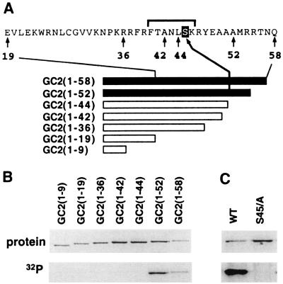
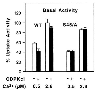
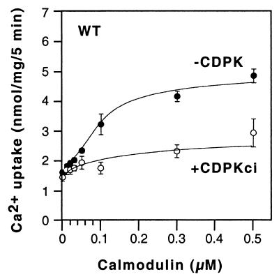
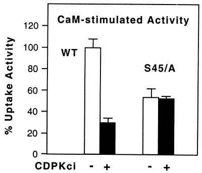
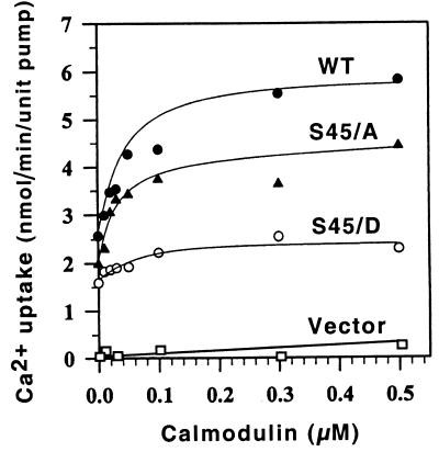
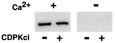
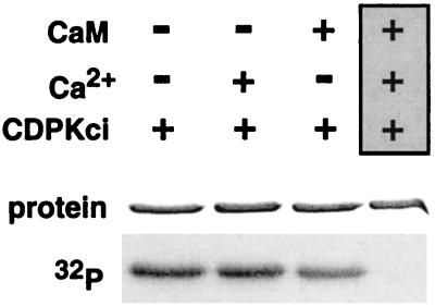
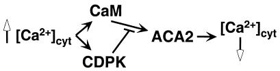
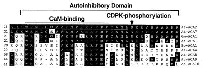
The magnitude and duration of a cytosolic Ca(2+) release can potentially be altered by changing the rate of Ca(2+) efflux. In plant cells, Ca(2+) efflux from the cytoplasm is mediated by H(+)/Ca(2+)-antiporters and two types of Ca(2+)-ATPases. ACA2 was recently identified as a calmodulin-regulated Ca(2+)-pump located in the endoplasmic reticulum. Here, we show that phosphorylation of its N-terminal regulatory domain by a Ca(2+)-dependent protein kinase (CDPK isoform CPK1), inhibits both basal activity ( approximately 10%) and calmodulin stimulation ( approximately 75%), as shown by Ca(2+)-transport assays with recombinant enzyme expressed in yeast. A CDPK phosphorylation site was mapped to Ser(45) near a calmodulin binding site, using a fusion protein containing the N-terminal domain as an in vitro substrate for a recombinant CPK1. In a full-length enzyme, an Ala substitution for Ser(45) (S45/A) completely blocked the observed CDPK inhibition of both basal and calmodulin-stimulated activities. An Asp substitution (S45/D) mimicked phosphoinhibition, indicating that a negative charge at this position is sufficient to account for phosphoinhibition. Interestingly, prior binding of calmodulin blocked phosphorylation. This suggests that, once ACA2 binds calmodulin, its activation state becomes resistant to phosphoinhibition. These results support the hypothesis that ACA2 activity is regulated as the balance between the initial kinetics of calmodulin stimulation and CDPK inhibition, providing an example in plants for a potential point of crosstalk between two different Ca(2+)-signaling pathways.


Figures

Figure 1
Figure 1
N-terminal domain of ACA2–1 is phosphorylated by CDPK at Ser45. (A) Diagram of deletions used to map a CDPK phosphorylation site. The sequence is shown surrounding the phosphorylation sited mapped to Ser45. The solid black lines indicate fusion proteins that were phosphorylated (shown in B). The bracket above the sequence identifies the tryptic peptide detected as a phosphopeptide of mass 860.4 by mass spectrometry analysis. (B) Phosphorylation of fusion proteins showing GC2(1–52) as the smallest fusion labeled by 32P. Row labeled “Protein”: GST-fusion proteins corresponding to different lengths of the N-terminal domain are shown by Coomassie stain. Protein names indicate which ACA2 residues are present. For example, GC2(1–9) indicates GST fusion of Ca2+ pump ACA2 residues 1–9. All fusion proteins have an N-terminal GST, a fragment of ACA2, and a C-terminal GFP (green fluorescent protein) as described (6). Row labeled “32P” shows the phosphorylation detected as 32P-labeling during a kinase reaction. Purified fusion proteins (≈2 μg) were incubated with 90 ng of a CPK1 mutant, KJM23-6H2, and 50 μM ATP spiked with [γ-32P]ATP for 60 min at 22°C in standard kinase reaction buffer without Ca2+. The proteins were analyzed by SDS/PAGE and autoradiography. (C) A substitution of Ser45 to Ala confirms the identification of a CPK phosphorylation site at Ser45. Rows correspond to markings in B. The protein marked WT (wild-type) contains the first 52 residues of the N terminus, GC2(1–52) from A. The protein marked S45/A is encoded by pACA2-N-S45/A, which is a derivative of GC2(1–52) with an Ala substitution for Ser45. Both proteins were subjected to a phosphorylation reaction as described in A. Proteins were then analyzed by SDS/PAGE and exposed to film. As a control to ensure that the kinase was active in all reactions, gels were overexposed to detect kinase autophosphorylation (not shown). Phosphorylation reactions were repeated two times with equivalent results.
Figure 2
Figure 2
Phosphorylation decreases the basal activity (without calmodulin) of wild-type ACA2, but not the mutant with an S45/A mutation. Membranes were isolated from yeast expressing a wild-type ACA2–1 (WT) or a mutant ACA2–3 (S45/A) harboring the S45/A substitution. Membranes were incubated with (+) or without (−) a Ca2+-independent CPK1 mutant, ΔNC31, in the absence of Ca2+. The transport assay contained 100 μM EGTA and 50 or 100 μM Ca2+ to give final [Ca2+] of 0.5 or 2.6 μM, respectively. Activity was calculated as net vanadate-sensitive Ca2+ transport during the first 5 min and expressed as a percent of wild type (100% = 1.7 nmol/5 min per mg of membrane). The activity of the mutant was normalized to the equivalent amount of wild-type enzyme. Relative levels of wild type and mutant proteins were estimated by immunoblots. The average activity (±SD) from two independent experiments is shown.
Figure 3
Figure 3
Calmodulin stimulation of ACA2 is decreased by a CDPK phosphorylation. Membranes isolated from yeast expressing ACA2–1 were incubated with ΔNC31 kinase as in Fig. 2. Net Ca2+ uptake (after 5 min) was determined by adding the CDPK-treated membranes to reaction mixtures containing 0–0.5 μM calmodulin. The average (±SD) from three independent determinations of two different membrane preparations is shown.
Figure 4
Figure 4
Phosphoinhibition of calmodulin stimulation is prevented by an S45/A mutation in ACA2. Membranes isolated from yeast expressing either ACA2–1 (WT) or ACA2–3 (S45/A) were incubated with kinase as described in Fig. 2. Net uptake (after 5 min) was determined with or without calmodulin (0.5 μM) in reaction mixtures containing 2.6 μM free Ca2+. Activity is presented as a percent of calmodulin-stimulated activity of wild-type (100% = 2.6 nmol/5 min per mg). The activity of the mutant enzyme was normalized to the equivalent amount of wild-type enzyme. The average (±SD) from two independent assays is shown. Assays on a second independent set of membrane preparations gave similar results.
Figure 5
Figure 5
The S45/D mutant is only weakly activated by calmodulin. Membranes isolated from yeast expressing ACA2–1 (WT), ACA2–3 (S45/A), ACA2–4 (S45/D), or vector only were used to determine the initial rate of uptake (after first 40 s) as a function of calmodulin at a free [Ca2+] of 2.6 μM. The maximum initial activity of the wild-type enzyme was 5.8 nmol/min per mg. The activity of mutant enzymes was normalized to the same amount of wild-type enzyme. Average of three independent assays is shown.
Figure 6
Figure 6
Phosphorylation of Ser45 does not disrupt calmodulin binding to the N-terminal domain. Fusion protein MC2-1-72 (0.3–3 μg) containing the first 72 residues of ACA2 was subjected to phosphorylation by a reaction with 90 ng of a Ca2+-independent CDPK mutant [CDPKci; either KJM23-6H2 (18) or ΔNC-31 (19)], and 300 μM ATP for 60 min at 22°C in a standard kinase reaction buffer without Ca2+ (18). The proteins were then tested for changes in calmodulin binding, as determined by gel overlay analysis in the presence (+) or absence (−) of 0.5 mM Ca2+, as described in ref. . Bound calmodulin was detected by enhanced chemiluminescence and exposure to x-ray film. One example is shown of three independent experiments. Equivalent results were obtained by using 6 or 60 nM biotinylated bovine calmodulin.
Figure 7
Figure 7
Phosphorylation of Ser45 is blocked by calmodulin binding. Fusion protein MC2-1-72 (2.5 μg) was preincubated in standard kinase reaction buffer with or without Ca2+ (100 μM) and calmodulin (CaM, 5 μM) for 15 min. Kinase reactions were started by adding 90 ng of a Ca2+-indpendent CDPK mutant, KJM23-6H2, and 50 μM ATP spiked with [γ-32P]ATP. Reactions were incubated for 60 min at 22°C. The proteins were subjected to SDS/PAGE and exposed to x-ray film. Row marked “protein” shows fusion proteins detected by Coomassie stain. Row marked “32P” shows an autoradiogram detecting the level of phosphorylation.
Figure 8
Figure 8
Diagram illustrating the opposing activities of calmodulin (CaM) and CDPKs on the activity of Ca2+ pump ACA2.
Figure 9
Figure 9
Alignment of ACA2-like pumps showing that a regulatory phosphorylation site at position Ser45 is not present in all pumps. Sequences war taken from Geisler et al. (11) with the addition of Gm-ACA1 from Moo Je Cho and Woosik Chung (personal communication). There is evidence from studies on ACA2 for a calmodulin-binding site in the region identified immediately upstream of Ser45 (6) and an autoinhibitory sequence contained within residues 20–44 (white line) (10). The potential limits of a functional autoinhibitory domain are marked based on mutations found in this region that disrupt autoinhibition (unpublished observations).

References

    1. Bush D S. Annu Rev Plant Physiol Plant Mol Biol. 1995;46:95–122.
    1. Sanders D, Brownlee C, Harper J F. Plant Cell. 1999;11:691–706. - PMC - PubMed
    1. Ghosh A, Greenberg M E. Science. 1995;268:239–247. - PubMed
    1. Axelsen K B, Palmgren M G. J Mol Evol. 1998;46:84–101. - PubMed
    1. Malmstrom S, Askerlund P, Palmgren M G. FEBS Lett. 1997;400:324–328. - PubMed

Publication types

MeSH terms