. 2000 Jun;20(12):4393-404.
doi: 10.1128/MCB.20.12.4393-4404.2000.

DNA repair protein Rad55 is a terminal substrate of the DNA damage checkpoints

Affiliations

DNA repair protein Rad55 is a terminal substrate of the DNA damage checkpoints

V I Bashkirov et al. Mol Cell Biol. 2000 Jun.

Abstract

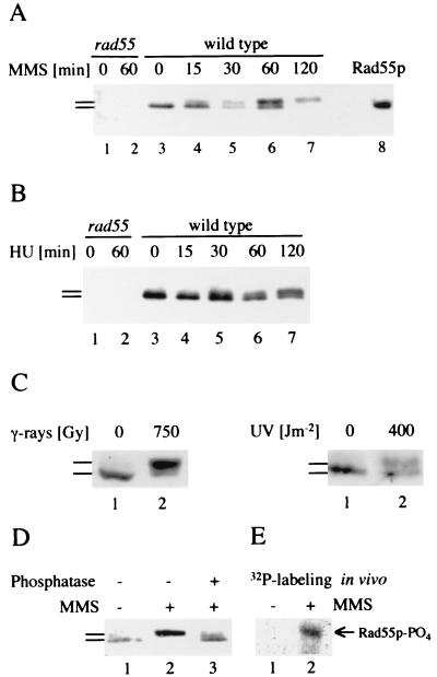
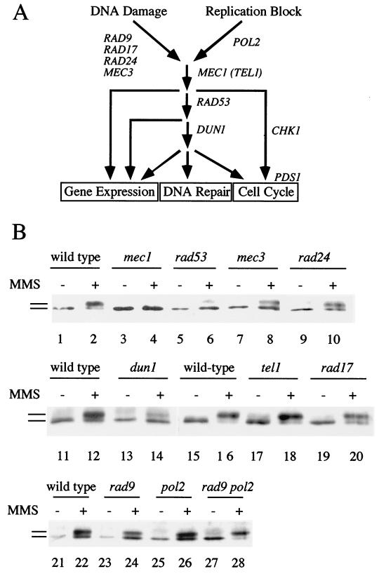
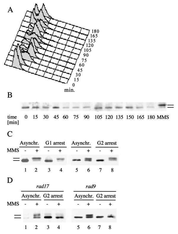
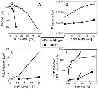
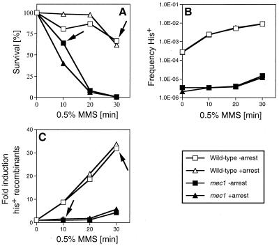
Checkpoints, which are integral to the cellular response to DNA damage, coordinate transient cell cycle arrest and the induced expression of DNA repair genes after genotoxic stress. DNA repair ensures cellular survival and genomic stability, utilizing a multipathway network. Here we report evidence that the two systems, DNA damage checkpoint control and DNA repair, are directly connected by demonstrating that the Rad55 double-strand break repair protein of the recombinational repair pathway is a terminal substrate of DNA damage and replication block checkpoints. Rad55p was specifically phosphorylated in response to DNA damage induced by the alkylating agent methyl methanesulfonate, dependent on an active DNA damage checkpoint. Rad55p modification was also observed after gamma ray and UV radiation. The rapid time course of phosphorylation and the recombination defects identified in checkpoint-deficient cells are consistent with a role of the DNA damage checkpoint in activating recombinational repair. Rad55p phosphorylation possibly affects the balance between different competing DNA repair pathways.


Figures

FIG. 1
FIG. 1
Phosphorylation of Rad55p in response to DNA damage and replication blocks. (A) Modification of Rad55p in response to DNA damage was analyzed by immunoprecipitation and immunoblotting. Lanes 1 and 2 contain precipitates from rad55Δ cells (WDHY1089) without MMS (lane 1) and after 60 min of exposure to 0.075% MMS (lane 2). Lanes 3 to 7 contain precipitates from wild-type cells (WDHY1075) without MMS (lane 3) and after increasing exposure time (15 to 120 min) to 0.075% MMS (lanes 4–7). In lane 8, extract of wild-type cells overexpressing Rad55p was directly blotted to show the position of the Rad55 protein. In lanes 5 and 7, accidentally less protein was loaded. (B) Modification of Rad55p in response to replication blocks. Cells were treated with HU and the Rad55p status was analyzed as in panel A. Lanes 1 and 2 contain precipitates from rad55Δ cells without HU (lane 1) and after 60 min of exposure to 200 mM HU (lane 2). Lanes 3 to 7 contain precipitates from wild-type cells without HU (lane 3) and after increasing exposure time (15 to 120 min) to 200 mM HU (lanes 4 to 7). (C) Modification of Rad55p in response to UV and gamma radiation. Exponentially growing wild-type cells (FF18984) were irradiated with gamma rays (137Cs source at 8 Gy/min) or UV rays (254 nm) at the indicated doses or mock irradiated. Cell extracts were prepared 1 h postradiation and analyzed for their Rad55p status. (D) Phosphorylation of Rad55p in response to DNA damage. The Rad55p status was analyzed in wild-type cells as in panel A from cells grown in the absence (lane 1) and in the presence (lanes 2 and 3) of MMS (0.075% for 120 min). An immunoprecipitate of a sample from MMS-treated cells was incubated with phosphatase before immunoblot analysis (lane 3). (E) Rad55p is phosphorylated in vivo in response to MMS. Wild-type cells (WDHY1075) were metabolically labeled with 32P in the presence (0.1%) and absence of MMS. Rad55p was immunoprecipitated and analyzed by autoradiography. Immunoblotting confirmed the position of unphosphorylated and phosphorylated Rad55p. Bars refer to the different forms of Rad55p.
FIG. 2
FIG. 2
Genetic control of Rad55p phosphorylation in response to DNA damage. (A) Overview of the DNA damage and replication block checkpoint in S. cerevisiae and the proposed functions of the genes used in panel B (18, 24, 68). DNA damage in G1 and G2 cells is sensed and/or processed by Rad9p, Rad17p, Rad24p, and Mec3p. Replication blocks are sensed by the DNA polymerase ɛ (Pol2p) (57). Both branches feed into the Mec1p kinase, which controls activation of the Rad53p kinase (69, 76). Rad53p controls some but not all checkpoint responses (12) and leads to the activation of the Dun1p kinase (93). Dun1p kinase is involved in the activation of some DNA damage-inducible genes (93) and, in one pathway with Rad53p, in G2/M cell cycle arrest parallel to a pathway acting through Chk1p kinase and Pds1p (24, 68). As shown here, Dun1p kinase is required for full phosphorylation of Rad55p in response to DNA damage (labeled DNA repair). (B) Rad55p phosphorylation in cycling cells depends on some but not all DNA damage checkpoint functions. Cells (0 or 90 min in 0.1% MMS at 24°C) were analyzed as described in the legend to Fig. 1; wild-type (TWY12; lanes 1 and 2), mec1-1 (TWY308; lanes 3 and 4), rad53 (TWY312; lanes 5 and 6), mec3-1 (TWY316; lanes 7 and 8), rad24-1 (TWY399; lanes 9 and 10), wild-type (Y300; lanes 11, 12, 15, 16, 21, and 22), dun1-Δ100 (Y286; lanes 13 and 14), tel1Δ (WDHY1227; lanes 17 and 18), rad17Δ (WDHY1234; lanes 19 and 20), rad9 (Y438; lanes 23 and 24), pol2-12 (Y439; lanes 25 and 26), and rad9 pol2-12 (Y440; lanes 27 and 28). The wild-type control is shown for each individual experiment. Bars refer to the different forms of Rad55p.
FIG. 3
FIG. 3
Rad55p phosphorylation in response to DNA damage during the cell cycle. (A) FACS analysis of a synchronized cell culture after α-factor arrest and release. At the indicated time intervals, aliquots were withdrawn, stained with propidium iodide, and analyzed by FACS. (B) Rad55p is not detectably phosphorylated during a normal cell cycle. At the same time intervals as in panel A, the Rad55p phosphorylation status was determined as described in the legend to Fig. 1. The rightmost lane (labeled MMS) shows a positive control (2 h with 0.1% MMS), indicating the migration behavior of phosphorylated Rad55p. (C) Rad55p phosphorylation in G1- and G2-arrested cells. Wild-type cells (FF181268) arrested either in G1 by α-factor (lanes 3 and 4) or in G2 by nocodazole (lanes 7 and 8) were treated with 0.1% MMS for 2 h (lanes 4 and 8) or left untreated (lanes 3 and 7). The Rad55p status was analyzed as described in the legend to Fig. 1. As a control, cycling wild-type cells were analyzed before (lanes 1 and 5) and after (lanes 2 and 6) MMS exposure. Asynchr., asynchronous. (D) Rad55p phosphorylation in G2-arrested cells is fully dependent on RAD9 or RAD17. The Rad55p status was analyzed as in panel A in rad9Δ cells (FF181270) left asynchronous (Asynchr.) (lanes 1 and 2) or arrested in G2 by nocodazole (lanes 3 and 4) and in rad17Δ cells (WDHY1236) left asynchronous (lanes 5 and 6) or arrested in G2 by nocodazole (lanes 7 and 8) after treatment with 0.1% MMS (lanes 2, 4, 6, and 8) or without MMS (lanes 1, 3, 5, and 7). Bars refer to the different forms of Rad55p.
FIG. 4
FIG. 4
mec1 cells are defective in DNA damage-induced mitotic recombination. (A) Survival of wild-type cells and mec1 cells after acute exposure to 0.5% MMS for the indicated times was measured as described elsewhere (65). (B) Absolute frequencies of Leu+ recombinants per viable cell with respect to MMS dose in wild-type and mec1 cells. (C) Fold induction of Leu+ recombinants with respect to MMS dose. (A to C) Shown is one experiment typical of three performed. The decrease in induced recombination in mec1 cells was seen in every experiment. (D) Induction of Leu+ recombinants per viable cell with respect to survival after MMS exposure in wild-type and mec1 cells. Given are the means of three determinations and standard deviations (error bars). Where no bars appear, the standard deviations were smaller than the symbols used. The wild-type strain was P7BAB, and the mec1 strain was NR110AB. The two arrows in panel D indicate a data point of similar survival between wild-type and mec1 cells (see text).
FIG. 5
FIG. 5
Artificial cell cycle arrest does not rescue the damage-induced recombination defect in mec1 cells. (A) Survival of wild-type cells and mec1 cells after acute exposure to 0.5% MMS for the indicated times was measured as described elsewhere (65). (B) Absolute frequencies of His+ recombinants per viable cell with respect to MMS dose in wild-type and mec1 cells. The spontaneous frequency for His+ recombinants in wild-type cells was 3.35 × 10⁻⁴ without arrest and 2.67 × 10⁻⁴ with arrest, and the values in mec1 cells were 3.45 × 10⁻⁶ without arrest and 2.1 × 10⁻⁶ with arrest. (C) Fold induction of His+ recombinants with respect to MMS dose. (A to C) Shown is one experiment typical of three to five performed. The decrease in induced recombination in mec1 cells was seen in every experiment. The wild-type strain was P7BAB, and the mec1 strain WDHY1558. The arrows in panels A and C indicate the effect on induced recombination at comparable survival levels for both strains (see text).
FIG. 6
FIG. 6
Model of checkpoint regulation of pathway competition in DNA repair. In wild-type cells, several pathways compete for the repair of DNA damage like a DSB (36, 61). In S. cerevisiae, the damage is preferentially repaired by recombinational repair, resulting primarily in gene conversions which are rarely (0 to 20%) associated with a crossing over (for a detailed discussion see reference and references therein). This avoidance of crossing over may be related to the frequent nondisjunction observed for mitotic crossing-over products (11). Under genotoxic stress, Rad55p is essential for the recombinational repair pathway (36, 61). A defect in the checkpoint (mec1) fails to phosphorylate Rad55p in response to DNA damage, which we hypothesize will lead to decreased efficiency in Rad51p filament formation (see reference 79) and a less efficient recombinational repair pathway. Under these conditions, other pathways will contribute more noticeably. BIR results in a genetic outcome that resembles a crossing-over (7, 48, 55, 61). NHEJ will not lead to recombinants (36, 53, 61). Depicted is a diploid cell in G1 with two homologues carrying three heterozygous markers (aA/bB/cC).

References

    1. Aboussekhra A, Vialard J E, Morrison D E, Delatorreruiz M A, Cernakova L, Fabre F, Lowndes N F. A novel role for the budding yeast RAD9 checkpoint gene in DNA damage-dependent transcription. EMBO J. 1996;15:3912–3922. - PMC - PubMed
    1. Al-Khodairy F, Carr A M. DNA repair mutants defining G2 checkpoint pathways in Schizosaccharomyces pombe. EMBO J. 1992;11:1343–1350. - PMC - PubMed
    1. Allen J B, Zhou Z, Siede W, Friedberg E C, Elledge S J. The SAD1/RAD53 protein kinase controls multiple checkpoints and DNA damage-induced transcription in yeast. Genes Dev. 1994;8:2401–2415. - PubMed
    1. Baumann P, West S C. Role of the human RAD51 protein in homologous recombination and double-stranded break repair. Trends Biochem Sci. 1998;23:247–251. - PubMed
    1. Bentley N J, Holtzman D A, Flaggs G, Keegan K S, DeMaggio A, Ford J C, Hoekstra M, Carr A M. The Schizosaccharomyces pombe rad3 checkpoint gene. EMBO J. 1996;15:6641–6651. - PMC - PubMed

Publication types

LinkOut - more resources