. 2000 Jun 6;97(12):6493-8.
doi: 10.1073/pnas.100135197.

Suppressor of cytokine signaling-3 preferentially binds to the SHP-2-binding site on the shared cytokine receptor subunit gp130

Affiliations

Suppressor of cytokine signaling-3 preferentially binds to the SHP-2-binding site on the shared cytokine receptor subunit gp130

S E Nicholson et al. Proc Natl Acad Sci U S A. .

Abstract

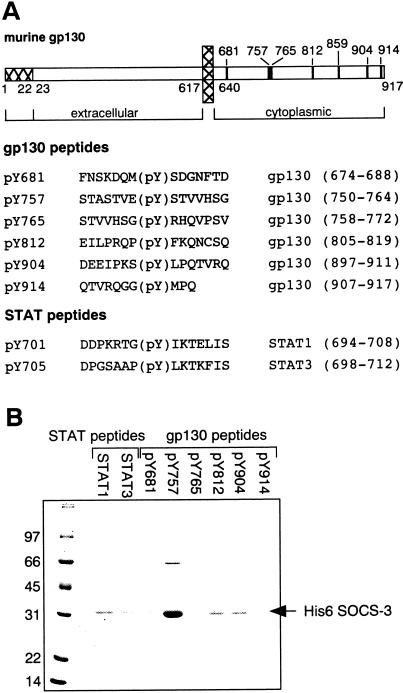
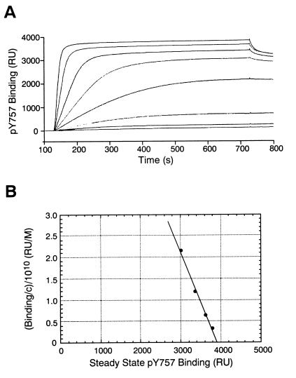
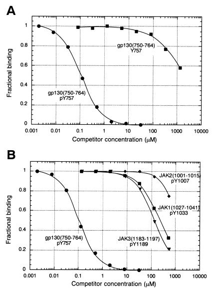
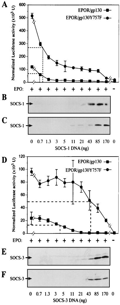
Suppressor of cytokine signaling-3 (SOCS-3) is one member of a family of intracellular inhibitors of signaling pathways initiated by cytokines that use, among others, the common receptor subunit gp130. The SH2 domain of SOCS-3 has been shown to be essential for this inhibitory activity, and we have used a quantitative binding analysis of SOCS-3 to synthetic phosphopeptides to map the potential sites of interaction of SOCS-3 with different components of the gp130 signaling pathway. The only high-affinity ligand found corresponded to the region of gp130 centered around phosphotyrosine-757 (pY757), previously shown to be a docking site for the tyrosine phosphatase SHP-2. By contrast, phosphopeptides corresponding to other regions within gp130, Janus kinase, or signal transducer and activator of transcription proteins bound to SOCS-3 with weak or undetectable affinity. The significance of pY757 in gp130 as a biologically relevant SOCS-3 docking site was investigated by using transfected 293T fibroblasts. Although SOCS-3 inhibited signaling in cells transfected with a chimeric receptor containing the wild-type gp130 intracellular domain, inhibition was considerably impaired for a receptor carrying a Y-->F point mutation at residue 757. Taken together, these data suggest that the mechanism by which SOCS-3 inhibits the gp130 signaling pathway depends on recruitment to the phosphorylated gp130 receptor, and that some of the negative regulatory roles previously attributed to the phosphatase SHP-2 might in fact be caused by the action of SOCS-3.


Figures

Figure 1
Figure 1
A gp130-derived phosphopeptide interacts specifically with SOCS-3. (A) Biotinylated phosphopeptides were synthesized corresponding to the regions of murine gp130 surrounding each cytoplasmic tyrosine residue. The pY812 peptide contained a cysteine residue in the reduced form, whereas a pY859 peptide was not synthesized, as this tyrosine is not conserved in human gp130. Similarly, peptides corresponding to regions surrounding the known tyrosine phosphorylation sites in STAT1 and STAT3 were also synthesized. (B) Peptides shown in A were immobilized on streptavidin–agarose resin and incubated with recombinant SOCS-3. Only the gp130 pY757 peptide showed any significant binding of SOCS-3 as detected by Coomassie-stained SDS/PAGE analysis of the resin eluates.
Figure 2
Figure 2
Functional characterization of SOCS-3 protein. (A) Biosensor analysis of SOCS-3 binding to the gp130-derived phosphopeptide biotin-STASTVEpYSTVVHSG. Sensorgrams correspond to a 2-fold serial dilution series of SOCS-3 (8.65–1,110 nM) binding to the immobilized peptide on a streptavidin sensorchip. (B) Determination of binding constants by Scatchard-type analysis. Data shown in A were used to calculate the association constant for SOCS-3 binding to the immobilized gp130 peptide. Plateau binding values at steady state (four highest concentrations of SOCS-3) were plotted against the ratio of plateau binding to SOCS-3 concentration (Binding/c). The association binding constant (Ka = 2.4 × 10⁷ M−1) was calculated from the slope of the plot of Binding/c vs. Binding. The dissociation constant (Kd) = 1/Ka = 42 nM.
Figure 3
Figure 3
Comparison of SOCS-3 affinity for phosphorylated vs. nonphosphorylated gp130 peptide and JAK-derived peptides. (A) Solution binding of phosphorylated and nonphosphorylated forms of gp130(750–764) peptide to SOCS-3 was measured in a competitive binding assay. The IC50s for inhibition of SOCS-3 binding to immobilized ligand were 110 ± 2.7 nM for the phosphorylated peptide and 2.1 ± 0.2 mM for the unphosphorylated peptide. (B) Solution binding of JAK-derived peptides to SOCS-3. These phosphopeptides represent the activation loop sequences in JAK1, 2, and 3 and contain a phosphotyrosine residue corresponding to the autophosphorylation site. The amino acid sequences of these peptides were JAK1: IETDKE(pY)YTVKDDRD, JAK2: LPQDKE(pY)YKVKEPGE, JAK3: LPLGKD(pY)YVVREPGQ. The IC50 values for these peptides were: JAK1, 230 ± 6.6 μM; JAK2, 1,200 ± 50 μM; JAK3, 140 ± 4.8 μM. The IC50 value for the gp130(750–764) phosphopeptide, measured in the same experiment, was 110 ± 4.6 nM
Figure 4
Figure 4
SOCS-3, but not SOCS-1, inhibition of gp130 signaling is mediated through gp130 Y757. (A) 293T cells were transiently transfected with cDNAs expressing SOCS-1 and either EPOR/gp130 or EPOR/gp130Y757F in the presence of the APRE-luc and Srα-β-gal reporter genes. Cells were incubated in the presence (+) or absence (−) of 10 units/ml hEPO overnight and cell extracts prepared. Luciferase activity from triplicate samples was determined and normalized against β-gal activity. (B) SOCS-1 protein levels in 293T cells expressing EPOR/gp130 were determined by Western blot with anti-Flag antibody. (C) SOCS-1 protein levels in 293T cells expressing EPOR/gp130Y757F. (D) 293T cells were transiently transfected with cDNAs expressing SOCS-3 and either EPOR/gp130 or EPOR/gp130Y757F in the presence of the APRE-luc and Srα-β-gal reporter genes. (E) SOCS-3 protein levels in 293T cells expressing EPOR/gp130 were determined by Western blot with anti-Flag antibody. (F) SOCS-3 protein levels in 293T cells expressing EPOR/gp130Y757F.

References

    1. Leonard W J, O'Shea J J. Annu Rev Immunol. 1998;16:293–322. - PubMed
    1. Hilton D J. Cell Mol Life Sci. 1999;55:1568–1577. - PMC - PubMed
    1. Hilton D J, Richardson R T, Alexander W S, Viney E M, Willson T A, Sprigg N S, Starr R, Nicholson S E, Metcalf D, Nicola N A. Proc Natl Acad Sci USA. 1998;95:114–119. - PMC - PubMed
    1. Kamura T, Sato S, Haque D, Liu L, Kaelin W G, Jr, Conaway R C, Conaway J W. Genes Dev. 1998;12:3872–3881. - PMC - PubMed
    1. Zhang J G, Farley A, Nicholson S E, Willson T A, Zugaro L M, Simpson R J, Moritz R L, Cary D, Richardson R, Hausmann G, et al. Proc Natl Acad Sci USA. 1999;96:2071–2076. - PMC - PubMed

Publication types

MeSH terms

Substances

LinkOut - more resources