. 2000 Jun 6;97(12):6809-14.
doi: 10.1073/pnas.120166397.

Severe deficiencies in dopamine signaling in presymptomatic Huntington's disease mice

Affiliations

Severe deficiencies in dopamine signaling in presymptomatic Huntington's disease mice

J A Bibb et al. Proc Natl Acad Sci U S A. .

Abstract

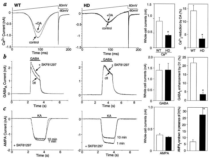
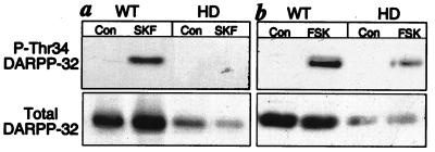
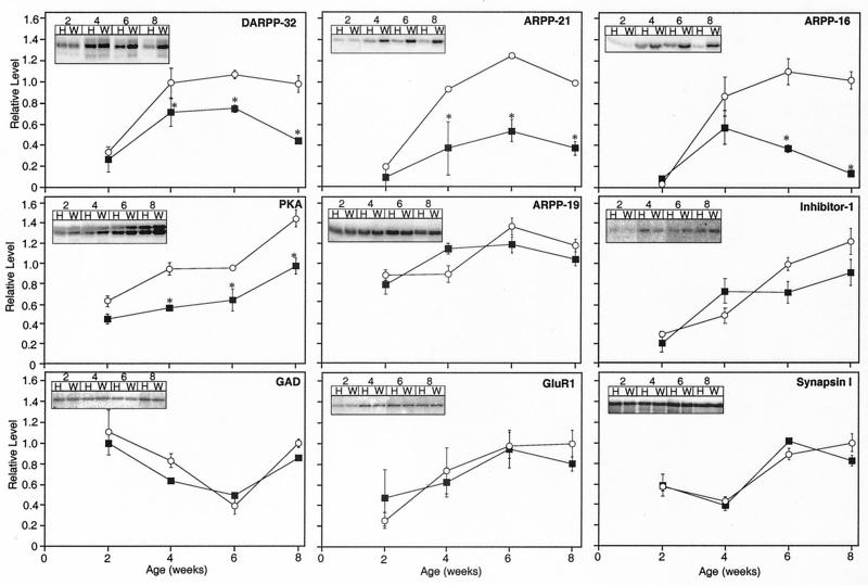
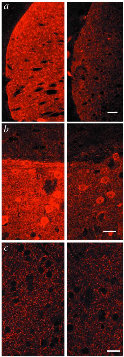
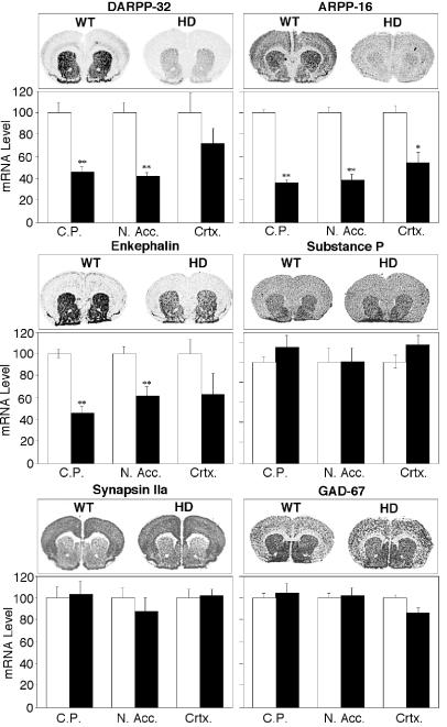
In Huntington's disease (HD), mutation of huntingtin causes selective neurodegeneration of dopaminoceptive striatal medium spiny neurons. Transgenic HD model mice that express a portion of the disease-causing form of human huntingtin develop a behavioral phenotype that suggests dysfunction of dopaminergic neurotransmission. Here we show that presymtomatic mice have severe deficiencies in dopamine signaling in the striatum. These include selective reductions in total levels of dopamine- and cAMP-regulated phosphoprotein, M(r) 32 kDA (DARPP-32) and other dopamine-regulated phosphoprotein markers of medium spiny neurons. HD mice also show defects in dopamine-regulated ion channels and in the D(1) dopamine/DARPP-32 signaling cascade. These presymptomatic defects may contribute to HD pathology.


Figures

Figure 1
Figure 1
Comparison of electrophysiological properties of WT and HD mice. Dopamine effects on voltage-gated Ca2+ current (a), GABAA current (b), and AMPA current (c) . To the right of each are summary histograms of the peak amplitudes of whole-cell currents (Left) and of the percent change induced by dopamine (DA) or the D1-class receptor agonist SKF81297 (Right) in WT (□) and HD mice (■). Data represent means ± SEM for n = 4–9; ∗, P < 0.05 compared with WT.
Figure 2
Figure 2
Comparison of DARPP-32 phosphorylation in response to a D1 agonist and forskolin in WT and HD mice. Striatal slices from 6-week-old mice were treated with either a dopamine D1 receptor agonist (a), SKF81297 (SKF), or an activator of adenylyl cyclase (b), forskolin (FSK). Homogenates of the slices were subjected to SDS/PAGE, proteins were transferred to polyvinylidene difluoride membranes, and phospho-Thr34 DARPP-32 was analyzed by immunoblotting using a phosphorylation state-specific antibody. Blots were reprobed to determine total levels of DARPP-32 (Lower).
Figure 3
Figure 3
Comparison of the levels of striatal proteins in WT and HD mice. Striatal homogenates of WT control (○) and HD (■) mice were subjected to SDS/PAGE and examined by immunoblotting with an antibody specific for the protein indicated. Mice were analyzed at 2, 4, 6, and 8 weeks of age. (Insets) Radiographic images showing representative protein bands detected in homogenates of HD (H) and WT (W) mice for each time point. Data represent means ± SEM for n = 6; ∗, P < 0.05 compared with WT.
Figure 4
Figure 4
Comparison of immunocytochemical staining for DARPP-32 in WT and HD mice. Striatal tissue of WT (Left) and HD mice (Right) was immunostained for DARPP-32 and GAD, and fluorescent images were collected by confocal laser scanning microscopy. (a) Image of coronal sections showing immunoreactivity for DARPP-32 in somata and dendrites of caudatoputamen. (b) Morphology of DARPP-32-positive medium spiny neurons and neuropil architectonics appear to be unaffected in the HD mouse. Intensely staining caudatoputamen (lower portion) contrasts with weakly staining cortex (upper portion). Note reduction in signal intensity in HD tissue. (c) Staining of striatal tissue by GAD-6 antibodies at ×630 magnification shows no differences between WT and HD model mice. (Scale bars in a, b, and c represent 250, 25, and 25 μm, respectively.)
Figure 5
Figure 5
Comparison of the levels of striatal gene expression in WT and HD mice. In situ hybridizations with probes specific for the detection of DARPP-32, ARPP-16, enkephalin, substance P, GAD-67, and synapsin IIa are shown. Representative labelings of coronal sections are shown for WT and HD mice. Histograms summarize quantification of signals in the caudatoputaman (C.P.), nucleus accumbens (N. Acc.) and cortex (Crtx) for WT (□) and HD mice (■). Data represent means ± SEM for n = 6; ∗∗, P < 0.001 and ∗, P < 0.01 compared with WT.

References

    1. Gusella J F, MacDonald M E. Semin Cell Biol. 1995;6:21–28. - PubMed
    1. Vonsattel J P, Myers R H, Stevens T J J R, Ferrante E D B, Richardson E P. J Neurol Exp Neuropathol. 1985;44:559–577. - PubMed
    1. Roos R A C. In: Handbook of Clinical Neurology. Vinke P J, Bruyn G W, Klawanm H L, editors. Vol. 49. Amsterdam: Elsevier; 1986. pp. 315–327.
    1. Huntington's Disease Collaborative Research Group. Cell. 1993;72:971–983. - PubMed
    1. Mangiarini L, Sathasivam K, Seller M, Cozens B, Harper A, Hetherington C, Lawton M, Trottier Y, Lehrach H, Davies S, Bates G P. Cell. 1996;87:493–506. - PubMed

Publication types