

Review
doi: 10.1083/jcb.149.5.995.
Syndecan-regulated receptor signaling
Affiliations
- PMID: 10831602
- PMCID: PMC2174822
- DOI: 10.1083/jcb.149.5.995
Item in Clipboard
Review
Syndecan-regulated receptor signaling
J Cell Biol.
.
No abstract available
Figures
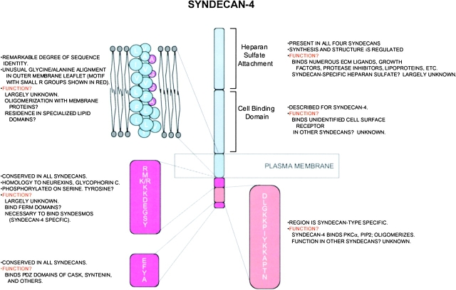
Syndecan functional domains. The extracellular, transmembrane and cytoplasmic domains of the syndecans contain important features, but the exact roles of these regions and how their function may be regulated remains uncertain.

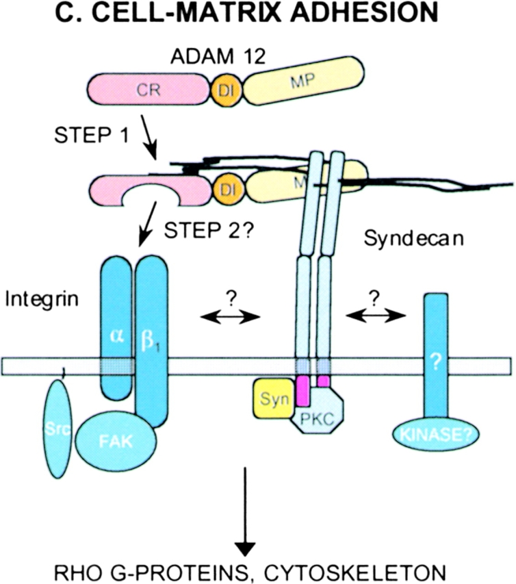
Syndecan-regulated signaling. Speculative examples of signaling mechanisms regulated by syndecans. (A) Cell–cell adhesion. Syndecan localized to sites of cell–cell adhesion (epithelial adherens junctions, neuronal synapses) may regulate the distribution of cytoskeletal/nuclear proteins CASK, protein 4.1 and β-catenin. (B) Signaling by HS-binding growth factors. Syndecan HS binds growth factors (GF) and growth factor receptors, regulating their assembly (positively or negatively) into signaling complexes. (C) Cell–matrix adhesion. Syndecans (syndecan-4) participate with integrins in focal adhesion assembly. Here, binding to ADAM 12 (step 1) may trigger syndecan core protein interactions with β1 integrins or unidentified signaling partners, leading to integrin activation. Alternatively, HS binding to the ADAM 12-cys region (CR) may alter CR domain conformation (step 2), exposing a cryptic binding site for β1 integrin binding and activation. This activation leads to focal adhesion and stress fiber formation, suggesting the participation of syndecan-4 and its associated syndesmos (Syn) and protein kinase C-α (PKC). DI, disintegrin domain; MP, metalloproteinase domain; FAK, focal adhesion kinase).

Syndecan-regulated signaling. Speculative examples of signaling mechanisms regulated by syndecans. (A) Cell–cell adhesion. Syndecan localized to sites of cell–cell adhesion (epithelial adherens junctions, neuronal synapses) may regulate the distribution of cytoskeletal/nuclear proteins CASK, protein 4.1 and β-catenin. (B) Signaling by HS-binding growth factors. Syndecan HS binds growth factors (GF) and growth factor receptors, regulating their assembly (positively or negatively) into signaling complexes. (C) Cell–matrix adhesion. Syndecans (syndecan-4) participate with integrins in focal adhesion assembly. Here, binding to ADAM 12 (step 1) may trigger syndecan core protein interactions with β1 integrins or unidentified signaling partners, leading to integrin activation. Alternatively, HS binding to the ADAM 12-cys region (CR) may alter CR domain conformation (step 2), exposing a cryptic binding site for β1 integrin binding and activation. This activation leads to focal adhesion and stress fiber formation, suggesting the participation of syndecan-4 and its associated syndesmos (Syn) and protein kinase C-α (PKC). DI, disintegrin domain; MP, metalloproteinase domain; FAK, focal adhesion kinase).
Syndecan-regulated signaling. Speculative examples of signaling mechanisms regulated by syndecans. (A) Cell–cell adhesion. Syndecan localized to sites of cell–cell adhesion (epithelial adherens junctions, neuronal synapses) may regulate the distribution of cytoskeletal/nuclear proteins CASK, protein 4.1 and β-catenin. (B) Signaling by HS-binding growth factors. Syndecan HS binds growth factors (GF) and growth factor receptors, regulating their assembly (positively or negatively) into signaling complexes. (C) Cell–matrix adhesion. Syndecans (syndecan-4) participate with integrins in focal adhesion assembly. Here, binding to ADAM 12 (step 1) may trigger syndecan core protein interactions with β1 integrins or unidentified signaling partners, leading to integrin activation. Alternatively, HS binding to the ADAM 12-cys region (CR) may alter CR domain conformation (step 2), exposing a cryptic binding site for β1 integrin binding and activation. This activation leads to focal adhesion and stress fiber formation, suggesting the participation of syndecan-4 and its associated syndesmos (Syn) and protein kinase C-α (PKC). DI, disintegrin domain; MP, metalloproteinase domain; FAK, focal adhesion kinase).
Comment on
-
The cysteine-rich domain of human ADAM 12 supports cell adhesion through syndecans and triggers signaling events that lead to beta1 integrin-dependent cell spreading.J Cell Biol. 2000 May 29;149(5):1143-56. doi: 10.1083/jcb.149.5.1143. J Cell Biol. 2000. PMID: 10831617 Free PMC article.
References
-
- Baciu P.C., Saoncella S., Lee S.H., Denhez F., Leuthardt D., Goetinck P.F. Syndesmos, a protein that interacts with the cytoplasmic domain of syndecan-4, mediates cell spreading and actin cytoskeletal organization. J. Cell Sci. 2000;113:315–324. - PubMed
-
- Bernfield M., Gotte M., Park P.W., Reizes O., Fitzgerald M.L., Lincecum J., Zako M. Functions of cell surface heparan sulfate proteoglycans. Annu. Rev. Biochem. 1999;68:729–777. - PubMed
-
- Cohen A.R., Woods D.F., Marfatia S.M., Walther Z., Chishti A.H., Anderson J.M., Wood D.F. Human CASK/LIN-2 binds syndecan-2 and protein 4.1 and localizes to the basolateral membrane of epithelial cells [published erratum appears in J. Cell Biol. 142:1157] J. Cell Biol. 1998;142:129–138. - PMC - PubMed
-
- Couchman J.R., Woods A. Syndecan-4 and integrinscombinatorial signaling in cell adhesion. J. Cell Sci. 1999;112:3415–3420. - PubMed
Publication types
MeSH terms
Substances
Grants and funding
LinkOut - more resources
Full Text Sources
Other Literature Sources
