. 2000 May 29;149(5):1143-56.
doi: 10.1083/jcb.149.5.1143.

The cysteine-rich domain of human ADAM 12 supports cell adhesion through syndecans and triggers signaling events that lead to beta1 integrin-dependent cell spreading

Affiliations

The cysteine-rich domain of human ADAM 12 supports cell adhesion through syndecans and triggers signaling events that lead to beta1 integrin-dependent cell spreading

K Iba et al. J Cell Biol. .

Abstract

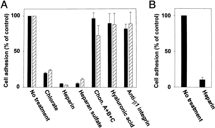
The ADAMs (a disintegrin and metalloprotease) family of proteins is involved in a variety of cellular interactions, including cell adhesion and ecto- domain shedding. Here we show that ADAM 12 binds to cell surface syndecans. Three forms of recombinant ADAM 12 were used in these experiments: the cys-teine-rich domain made in Escherichia coli (rADAM 12-cys), the disintegrin-like and cysteine-rich domain made in insect cells (rADAM 12-DC), and full-length human ADAM 12-S tagged with green fluorescent protein made in mammalian cells (rADAM 12-GFP). Mesenchymal cells specifically and in a dose-dependent manner attach to ADAM 12 via members of the syndecan family. After binding to syndecans, mesenchymal cells spread and form focal adhesions and actin stress fibers. Integrin beta1 was responsible for cell spreading because function-blocking monoclonal antibodies completely inhibited cell spreading, and chondroblasts lacking beta1 integrin attached but did not spread. These data suggest that mesenchymal cells use syndecans as the initial receptor for the ADAM 12 cysteine-rich domain-mediated cell adhesion, and then the beta1 integrin to induce cell spreading. Interestingly, carcinoma cells attached but did not spread on ADAM 12. However, spreading could be efficiently induced by the addition of either 1 mM Mn(2+) or the beta1 integrin-activating monoclonal antibody 12G10, suggesting that in these carcinoma cells, the ADAM 12-syndecan complex fails to modulate the function of beta1 integrin.


Figures

Figure 1
Figure 1
Mesenchymal cell attachment and spreading in response to rADAM 12-cys. (A) Cell attachment of MG-63 cells plated on rADAM 12-cys (20 μg/μl). Bar, 16 μm. (B) Dose-dependent cell attachment of SV-HFO (filled circles) and RD cells (open circles) plated on rADAM 12-cys. (C) Cell attachment of the SV-HFO cell line plated on rADAM 12-cys, rADAM 12-disintegrin, rADAM 12-metallo, rADAM 12-DC, laminin-1, and fibronectin (20 μg/ml). BSA represents the background value when no substrate was added. Ordinates represent OD values at 590 nm, and abscissa the concentration of the substrate in μg/ml as indicated. Each point represents the mean ± SEM of triplicate wells.
Figure 2
Figure 2
Focal adhesion formation and reorganization of the actin cytoskeleton in response to rADAM-cys. (A) SV-HFO cells plated on rADAM 12-cys and stained with mAbs to vinculin. Numerous distinct focal adhesions are seen. (B) SV-HFO cells plated on rADAM 12-cys and stained with phalloidin to detect F-actin. An elaborate organization of stress fibers is evident. Bars, 6 μm.
Figure 3
Figure 3
Cell attachment on rADAM 12-cys is dependent on cell surface heparan sulfate. (A) Cell attachment of SV-HFO cells (filled bars) and MRC-5 (hatched bars) plated on rADAM 12-cys. (B) Cell attachment of SV-HFO cells (filled bars) plated on rADAM 12-DC. The assays were performed in the presence or absence of the reagents indicated (chlorate 20 mM and the other reagents 10 μg/ml). Ordinates represent percentage adhesion. Each point represents the mean ± SEM of triplicate wells.
Figure 4
Figure 4
Cell attachment to rADAM 12-cys is mediated through the syndecans. (A) Cell attachment of ARH-77 cells transfected with either an empty vector (Neo), syndecan-1, -2, or -4 (Syn-1, -2, or -4), or with glypican-1 (Glyp-1) and plated on rADAM 12-cys (20 μg/μl) or in B, on rADAM 12-DC. (C) Cell attachment on rADAM 12-cys of syndecan-1–expressing cells treated with chlorate, heparin, heparan sulfate, chondroitin sulfate ABC (Chon A+B+C), heparitinase (Hep-ase) or chondroitinase ABC (Chon-ase ABC). ARH-77–TDM cells express syndecan-1 lacking heparan sulfate attachment sites but with intact chondroitin sulfate chains. (D) Cell attachment on rADAM 12-cys of ARH-77 cells expressing truncated syndecan-1 (Syn/279t) or chimeric proteoglycans (Syn/Glyp and Glyp/Syn). Each point represents the mean ± SEM of triplicate wells. Ordinates represent percentage of adhesion or OD values at 590 nm. Insets in D show syndecan-1 cell surface immunostaining of the ARH-77 Syn/Glyp cells (upper) and glypican-1 cell surface immunostaining of ARH-77 Glyp/Syn (lower). Bars, 22 μm.
Figure 5
Figure 5
Syndecan-4 binds to rADAM 12-cys. (A) RT-PCR demonstrates the expression of syndecan-1 (lane 2), -2 (lane 3), and -4 (lane 4) transcripts by the RD cells. Lane 1 is a negative control. (B) RD cell extracts were separated by DEAE Sephacel, treated with heparitinase and chondroitinase resolved by SDS-PAGE, and membranes probed with the pan-syndecan antibody that recognizes the conserved C2 region in the cytoplasmic domains of all the syndecan family members (lane 1) or a preimmune serum (lane 2). (C) SDS-PAGE of syndecan-4 from RD cells isolated by rADAM 12-cys affinity chromotography. Lane 1 shows a smeared band with an apparent M r 200,000. After treatment with heparitinase, a single distinct band with an apparent M r 35,000 (lane 2) is seen. A polyclonal antibody to syndecan-4 identified this band as syndecan-4 (lane 3). (D) Purified iodinated syndecan-4 before (lane 1) and after (lane 2) removal of the heparan sulfate glycosaminoglycan side chains by heparatinase treatment. (E) Solid phase binding assay demonstrating that syndecan-4 binds bFGF and rADAM 12-cys (filled bars) and that treatment of syndecan-4 with heparitinase completely inhibits its binding to rADAM 12-cys (hatched bars).
Figure 6
Figure 6
Full-length human ADAM 12 binds to syndecan-expressing cells. ARH-77 cells were incubated with ADAM 12-GFP–containing medium for 1 h at 4°C and examined by fluorescence microscopy. (A) Distinct cell surface fluorescence is observed along the cell surface of ARH-77 cells transfected with syndecan-1, but (B) no cell surface fluorescence is seen in control ARH-77 cells transfected with vector only. (C) Secreted GFP (not linked to ADAM 12) did not bind to syndecan-1–expressing cells, and (D) no fluorescence was observed when the cells had been pretreated with heparitinase. Phase contrast of the control ARH-77 neo cells examined in B is shown in E. Bars, 20 μm.
Figure 8
Figure 8
β1 integrin–deficient chondroblasts attach but do not spread on rADAM 12-cys. (A and B) Cell attachment of β1 integrin chondroblasts (+c/+c) plated on rADAM 12-cys and laminin-1, respectively. (C and D) Cell attachment of β1 integrin−/− chondroblasts on rADAM 12-cys and laminin-1, respectively. Bars, 14 μm. (E) Chondroblast cell attachment (filled bars) and the effect of 10 μg/ml of heparin (hatched bars) on rADAM 12-cys or laminin-1 as a substrate. The substrate concentration was 20 μg/ml. Ordinate represents OD values at 590 nm. (F) FACS analysis confirming the absence of β1 integrin on −/− chondroblasts.
Figure 7
Figure 7
Cell spreading in response to rADAM 12-cys is mediated by β1 integrin. (A) Cell attachment of SV-HFO cells with the characteristic spread morphology is shown. (B–D) In the presence of function-blocking mAbs to β1 integrin (AIIB2), the cells attached but no longer spread, and displayed a rounded morphology with many cytoplasmic projections. Three representative fields are shown. Bars: 10 μm (A, B, and D); and 12 μm (C).
Figure 9
Figure 9
Multiple integrins are involved in rADAM 12-cys–mediated cell spreading. (A) MRC-5 cells plated on rADAM 12-cys had a spread and flattened morphology. (B) In contrast, cells treated with function-blocking mAbs to α3 + α5 + α6 integrins attached but did not spread. Note the rounded cell morphology with numerous cytoplasmic projections. Bars: 22 μm (A); and 26 μm (B).
Figure 10
Figure 10
Carcinoma cells can be induced to spread by activating the β1 integrins. RKO colon carcinoma cells were allowed to attach to laminin (A and B), rADAM 12-cys (C–F), or rADAM 12-DC (G and H) for 30 min followed by the addition of reagents known to stimulate β1 integrin activity. RKO cells were plated on laminin either in the absence (A) or in the presence (B) of 1 mM Mn2+. RKO cells were plated on rADAM 12-cys in the absence (C), or presence (D) of 1 mM Mn2+, or in the presence of control β1 integrin mAb K20 (10 μg/ml) (E), or the β1 integrin–activating mAb 12G10 (10 μg/ml) (F). Similarly RKO cells were plated on rADAM 12-DC in the absence (G) or presence (H) of 1 mM Mn2+. Bar, 12 μm. (I) RKO cell attachment (filled bars) and the effect of 1 mM Mn2+ (hatched bars) on rADAM 12-cys, rADAM 12-DC, laminin-1, or fibronectin as substrates (20 μg/ml). Ordinate represents OD values at 590 nm.

Comment in

  • Syndecan-regulated receptor signaling.
    Rapraeger AC. Rapraeger AC. J Cell Biol. 2000 May 29;149(5):995-8. doi: 10.1083/jcb.149.5.995. J Cell Biol. 2000. PMID: 10831602 Free PMC article. Review. No abstract available.

Similar articles

Cited by

References

    1. Allen R.E., Dodson M.V., Luiten L.S. Regulation of skeletal muscle satellite cell proliferation by bovine pituitary fibroblast growth factor. Exp. Cell Res. 1984;152:154–160. - PubMed
    1. Almeida E.A., Huovila A.P., Sutherland A.E., Stephens L.E., Calarco P.G., Shaw L.M., Mercurio A.M., Sonnenberg A., Primakoff P., Myles D.G. Mouse egg integrin alpha 6 beta 1 functions as a sperm receptor. Cell. 1995;81:1095–1104. - PubMed
    1. Anton M., Graham F.L. Site-specific recombination mediated by an adenovirus vector expressing the Cre recombinase proteina molecular switch for control of gene expression. J. Virol. 1995;69:4600–4606. - PMC - PubMed
    1. Aszodi A., Pfeifer A., Ahmad M., Glauner M., Zhou X.H., Ny L., Andersson K.E., Kehrel B., Offermanns S., Fassler R. The vasodilator-stimulated phosphoprotein (VASP) is involved in cGMP- and cAMP-mediated inhibition of agonist-induced platelet aggregation, but is dispensable for smooth muscle function. EMBO (Eur. Mol. Biol. Organ.) J. 1999;18:37–48. - PMC - PubMed
    1. Baciu P.C., Goetinck P.F. Protein kinase C regulates the recruitment of syndecan-4 into focal contacts. Mol. Biol. Cell. 1995;6:1503–1513. - PMC - PubMed

Publication types

MeSH terms