. 2000 May 29;149(5):1157-66.
doi: 10.1083/jcb.149.5.1157.

Tissue plasminogen activator-mediated fibrinolysis protects against axonal degeneration and demyelination after sciatic nerve injury

Affiliations

Tissue plasminogen activator-mediated fibrinolysis protects against axonal degeneration and demyelination after sciatic nerve injury

K Akassoglou et al. J Cell Biol. .

Abstract

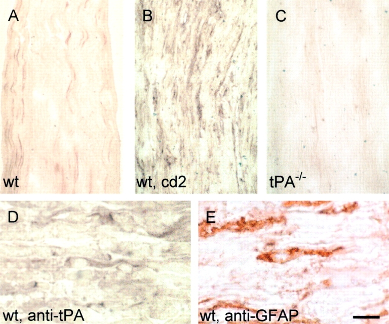
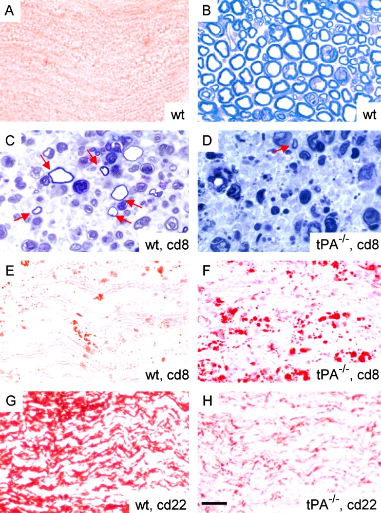
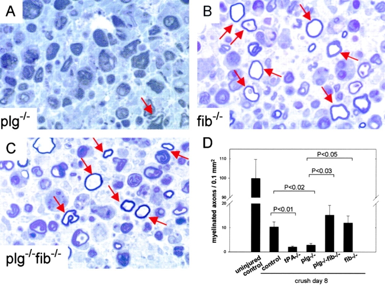
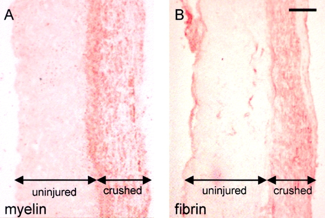
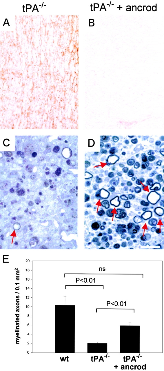
Tissue plasminogen activator (tPA) is a serine protease that converts plasminogen to plasmin and can trigger the degradation of extracellular matrix proteins. In the nervous system, under noninflammatory conditions, tPA contributes to excitotoxic neuronal death, probably through degradation of laminin. To evaluate the contribution of extracellular proteolysis in inflammatory neuronal degeneration, we performed sciatic nerve injury in mice. Proteolytic activity was increased in the nerve after injury, and this activity was primarily because of Schwann cell-produced tPA. To identify whether tPA release after nerve damage played a beneficial or deleterious role, we crushed the sciatic nerve of mice deficient for tPA. Axonal demyelination was exacerbated in the absence of tPA or plasminogen, indicating that tPA has a protective role in nerve injury, and that this protective effect is due to its proteolytic action on plasminogen. Axonal damage was correlated with increased fibrin(ogen) deposition, suggesting that this protein might play a role in neuronal injury. Consistent with this idea, the increased axonal degeneration phenotype in tPA- or plasminogen-deficient mice was ameliorated by genetic or pharmacological depletion of fibrinogen, identifying fibrin as the plasmin substrate in the nervous system under inflammatory axonal damage. This study shows that fibrin deposition exacerbates axonal injury, and that induction of an extracellular proteolytic cascade is a beneficial response of the tissue to remove fibrin. tPA/plasmin-mediated fibrinolysis may be a widespread protective mechanism in neuroinflammatory pathologies.


Figures

Figure 1
Figure 1
tPA is the major PA induced after sciatic nerve injury. In situ zymography to detect proteolytic activity 8 d after crush (cd8) showed a small proteolytic zone on wild-type uninjured sciatic nerve (A) and an increase of activity after injury (B, dark zone around nerve). C–E show nerves after injury. tPA−/− sciatic nerve (C) did not show any activity, whereas uPA−/− nerve (D) showed proteolytic activity similar to the wild type (B). Proteolytic activity of wild-type sciatic nerve was inhibited by tPA-STOP (E), a tPA inhibitor, but was unaffected by amiloride (F), a uPA inhibitor. Assay time was 6 h. Bar, 1 mm.
Figure 2
Figure 2
tPA is produced by Schwann cells after sciatic nerve crush. Immunocytochemistry with an antibody against tPA on longitudinal cryostat sections of wild-type, uninjured sciatic nerve revealed tPA staining only of the sciatic nerve vasculature (A). As early as 2 d after crush (cd2) tPA immunoreactivity was increased in the endoneurium (B). Sciatic nerve from a tPA−/− mouse showed no immunoreactivity with the tPA antibody (C). Immunostaining of parallel sections of a wild-type sciatic nerve 2 d after crush showed similar morphology between tPA immunoreactive cells (D) and GFAP-positive Schwann cells (E). The number of GFAP-positive cells was equivalent in wild-type and tPA−/− nerves before and after crush, suggesting that the number of Schwann cells is not significantly different in the two genotypes. Bar: (A–C) 93 μm; (D–E) 18 μm.
Figure 3
Figure 3
Axonal degeneration and demyelination are exacerbated in tPA-deficient mice after sciatic nerve crush. Oil Red O staining of cryostat sections of uninjured wild-type sciatic nerve (A) revealed normal myelin distribution, and toluidine blue staining of sciatic nerve semi-thin cross-sections showed normal axon morphology (B). 8 d after crush (cd8), toluidine blue staining of semi-thin cross-sections of wild-type mice (C) demonstrated more myelinated axons (arrows) than tPA−/− mice (D). Oil Red O staining showed increased accumulation of myelin and lipid debris in the tPA−/− (F) compared with the wild-type (E) sciatic nerve. 22 d after crush (cd22), staining for myelin basic protein revealed fewer myelinated axons in tPA−/− (H) than in wild-type (G) nerves. Bar, 18 μm.
Figure 4
Figure 4
tPA protects from axonal degeneration through a proteolytic mechanism. Toluidine blue staining of sciatic nerve semi-thin cross-sections of plg−/− mice (A) reveals exacerbated axonal damage. Fib−/− mice (B) and plg−/−fib−/− (C) mice showed myelinated axons similar in number to wild-type mice (C). (D) Quantification of myelinated axons. First column shows uninjured sciatic nerve (n = 4). After crush, tPA−/− (n = 6) and plg−/− (n = 4) nerves showed significantly fewer myelinated axons compared with control injured nerve (n = 5) (P < 0.01 and P < 0.04, respectively). Crushed fib−/− (n = 7) and plg−/−fib−/− (n = 4) nerves showed the same number of axons as control crushed nerves. Plg−/−fib−/− nerves showed more myelinated axons compared with tPA−/− nerves (P < 0.04). Uninjured sciatic nerves from all genotypes had similar number of myelinated axons and similar morphology. Data are expressed as means ± SEM. Statistical comparisons between medians were made with the t test. Scale as in Fig. 3.
Figure 5
Figure 5
Fibrin(ogen) deposition increases after sciatic nerve injury and correlates with axonal degeneration and demyelination. Staining of parallel sections of a partially crushed wild-type sciatic nerve 8 d after injury with Oil Red O (A), a myelin stain, and antifibrinogen antibody (B) revealed that the crushed part of the nerve, which underwent axonal degeneration (A, Oil Red O stained aggregates represent myelin debris accumulation), also had extensive deposition of fibrin(ogen) (B), whereas the immediately adjacent, uninjured region was free of fibrin(ogen). Double-headed arrows indicate uninjured and crushed regions. Bar, 165 μm.
Figure 6
Figure 6
Pharmacological depletion of fibrin(ogen) reduces axonal damage in tPA−/− mice. Immunostaining for fibrin(ogen) revealed deposition of fibrin(ogen) in tPA−/− (A) mice, whereas ancrod-treated tPA−/− (B) mice showed little fibrin(ogen) immunoreactivity. Toluidine blue staining of semi-thin cross-sections of crushed sciatic nerve (8 d after injury) of tPA−/− mice treated with ancrod showed an increase of myelinated axons (D) compared with buffer-treated tPA−/− mice (C). (E) Quantification of myelinated axons 8 d after sciatic nerve crush. tPA−/− mice treated with ancrod (n = 4) showed significantly more myelinated axons than buffer-treated tPA−/− mice (n = 6, P < 0.01). The difference between tPA−/− mice treated with ancrod and wild-type controls (n = 5) was not statistically significant (ns). Data are expressed as means ± SEM. Statistical comparisons between medians were made with the t test. Bar: (A and B) 113 μm; (C and D) 22 μm.
Figure 7
Figure 7
Depletion of fibrin(ogen) reduces muscle atrophy. Muscle atrophy 8 d after sciatic nerve crush was increased in the absence of tPA and decreased after fibrinogen depletion. Muscle mass in wild-type mice (n = 7) dropped 24.5 ± 2.7% compared with the unlesioned, contralateral side, whereas in tPA−/− mice (n = 7), muscle mass dropped 40.1 ± 3.8% (P < 0.005 compared with wild-type mice). After depletion of fibrinogen, muscle mass in tPA−/− mice treated with ancrod (n = 5) muscle mass dropped 16.6 ± 3.4% (P < 0.001 compared with tPA−/− mice). Data are expressed as means ± SEM. Statistical comparisons between medians were made with the t test.

References

    1. Akassoglou K., Probert L., Kontogeorgos G., Kollias G. Astrocyte-specific but not neuron-specific transmembrane TNF triggers inflammation and degeneration in the central nervous system of transgenic mice. J. Immunol. 1997;158:438–445. - PubMed
    1. Akassoglou K., Bauer J., Kassiotis G., Pasparakis M., Lassmann H., Kollias G., Probert L. Oligodendrocyte apoptosis and primary demyelination induced by local TNF/p55TNF receptor signaling in the central nervous system of transgenic micemodels for multiple sclerosis with primary oligodendrogliopathy. Am. J. Pathol. 1998;153:801–813. - PMC - PubMed
    1. Akenami F.O., Koskiniemi M., Farkkila M., Vaheri A. A. cerebrospinal fluid plasminogen activator inhibitor-1 in patients with neurological disease. J. Clin. Pathol. 1997;50:157–160. - PMC - PubMed
    1. Akenami F.O., Siren V., Wessman M., Koskiniemi M., Vaheri A. Tissue plasminogen activator gene expression in multiple sclerosis brain tissue. J. Neurol. Sci. 1999;165:71–76. - PubMed
    1. Arnold D.L. Magnetic resonance spectroscopyimaging axonal damage in MS. J. Neuroimmunol. 1999;98:2–6. - PubMed

Publication types

MeSH terms